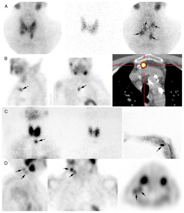
FIGURE 1.
Case examples of parathyroid scintigraphy in sHPT. A, Parathyroid scintigraphy prior to initial surgery for secondary HPT. Subtraction protocol: 123I at 2 hours post-administration of 12 MBq from 740 MBq of 99mTc-sestamibi at time 0, simultaneous dual-tracer image recording (planar + pinhole), digital subtraction of images (99mTc-sestamibi minus 123I). Four enlarged parathyroid glands are seen on pinhole subtraction images in this patient, with asymmetrical gland locations (arrows). B–D, Parathyroid scintigraphy for persistent or recurrent sHPT. B, Recurrent tertiary HPT related to a supernumerary ectopic gland (left and middle: SPECT images; right: fusion SPECT/CT image). C, Recurrence caused by supernumerary ectopic “upper mediastinal” parathyroid gland and forearm graft hyperplasia (arrows). Left: planar static cervicomediastinal 99mTc-sestaMIBI; middle: planar static cervicomediastinal 123I image; right: 99mTc-sestaMIBI planar image centered over the graft. D, Recurrent sHPT related to parathyromatosis. The patient had total thyroid ablation during one of the previous parathyroid surgeries. Multiple foci of 99mTc-sestaMIBI uptake, corresponding to hyperfunctioning parathyroid tissue are seen in upper lateral right neck.

