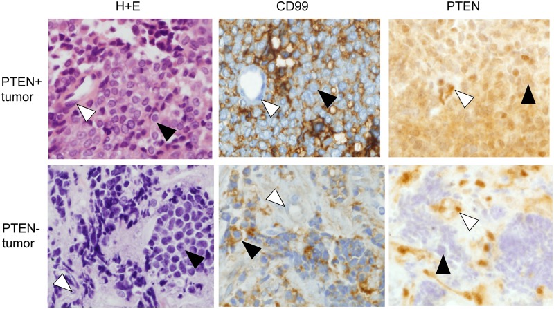
Figure 4. PTEN expression in patient tumor samples.
PTEN immunohistochemical staining in a representative PTEN-positive tumor and a PTEN-negative tumor. H+E histology and CD99 immunohistochemical staining are also shown. White arrowheads indicate representative vascular endothelial cells (strongly PTEN+ and CD99-); black arrowheads indicate representative tumor cells (CD99+). Note that vascular endothelial cells in both tumors show strong PTEN immunoreactivity of similar intensity (white arrowheads), but tumor cells only in the top tumor are PTEN immunoreactive (black arrowheads).

