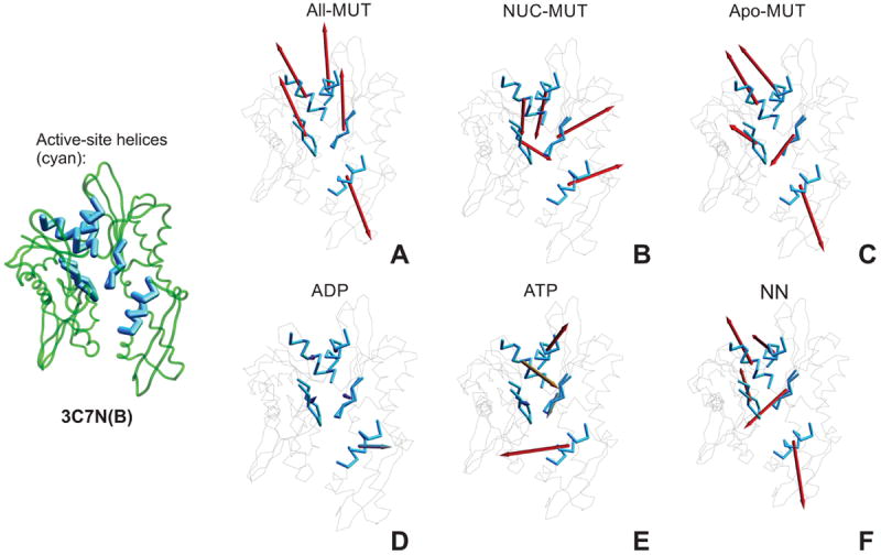
Figure 10.

Active-site fragment vectors, per vector class: A. All-mutual, B. NUC-mutual, C. NN- mutual, D. ADP-unique, E. ATP-unique class, and F. NN-unique. Each vector is shown as an arrow anchored in the center of the corresponding fragment, with direction and length corresponding to the direction and extent of the motion of this fragment follwing the respective eigenvector. The active site and its immediate periphery are shown in blue. The length of vectors reflects their magnitude, to a maximal cut-off. For clarity, the structure of the NBD with active-site components shown in thicker lines is included on the left to panels A – E.
