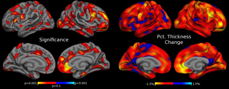
Figure 9. Cortical Thickness Estimates Correlate with Motion after extreme QC.
Regions of significant cortical thickness change associated with increased motion after removing scans that fail QC and scans with a warning. The p-value map (left) is not FDR thresholded (effects disappear after FDR). Compare to Figs. 8 and 4 and see description for details.

