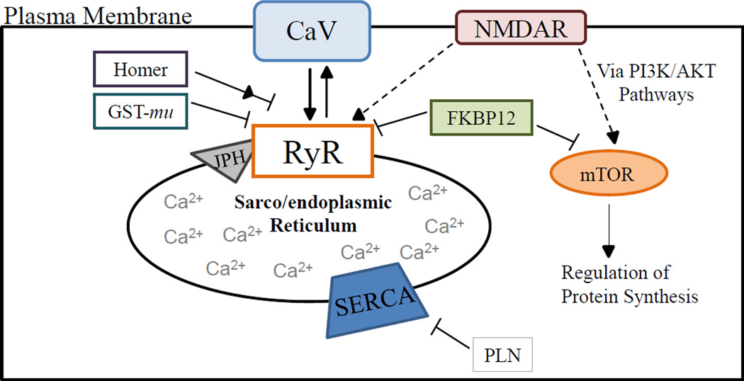
Figure 1.
Proteins involved in RyR Ca2+ signaling dynamics and potential RyR related signaling partners. Arrow heads and blunt ended lines represent activating or inhibiting regulation respectively. Full versus dashed lines represent an established versus a proposed relationship, respectively. Note that the diagram is not isoform or tissue specific but rather proteins displayed relate to Ca2+ signaling in skeletal muscle, cardiac muscle or brain tissue. Also, this is not an exhaustive list of RyR regulatory proteins or signaling partners. The Ca2+ channel interacts with or is regulated by numerous other plasma membrane bound signaling receptors, cytoplasmic accessory proteins and integral and luminal sarcoplasmic reticulum proteins not measured in the current study. Abbreviations see Table 1. Information gathered from (Pouliquin and Dulhunty, 2009; Van Dongen et al, 2009; Hoeffer and Klann, 2010; Pessah et al., 2010; Martín-Cano et al., 2013).

