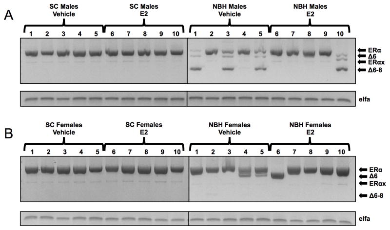
Figure 6. Expression of 3′-end ERα variants in the liver of individual male (A) and female (B) killifish after treatment with estradiol or vehicle alone, as determined by PCR analysis and agarose gel electrophoresis.
Adult killifish were injected with estradiol or vehicle alone (see legend to Figure 4) and livers processed to RNA. cDNA from individual male or female killifish livers (lanes 1–10) was amplified with primers in exon 5 (#5) and the untranslated region of exon 9 (#9) in order to generate full-length and possible 3′-end deletion variants of ERα at the same time. Products were size-separated on agarose gels: full-length, 806 bp; ERαΔ6, 668 bp; ERαx, 476 bp; ERαΔ6–8, 350 bp. RNA extraction, reverse transcription, polymerase, primer concentrations, cycling parameters, and agarose gel electrophoresis were exactly as described in Materials & Methods (sections 2.2 and 2.4).

