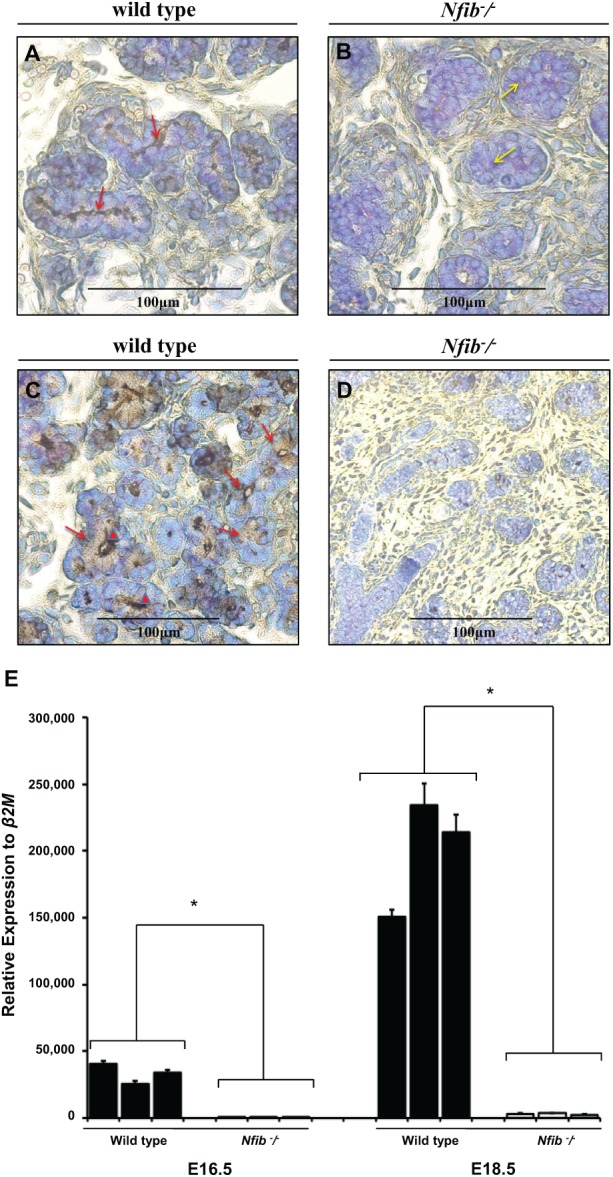
Figure 3.

The Nfib −/− mouse submandibular gland (SMG) at E18.5 lack aquaporin 5 and SMG protein C (SMGC) expression. Salivary glands were dissected, embedded in paraffin, and stained with aquaporin 5 or SMGC antibodies as described in the Materials and Methods. Strong aquaporin 5 immunohistochemical localization along the luminal membrane of the tubule secretory cells from wild-type mouse SMG was observed (A, red arrows). However, there was an absence of immunohistochemical staining for aquaporin 5 in the terminal buds of the Nfib −/− mouse SMG and failure to form lumens (or displayed poorly formed lumens) (B, yellow arrows). (B) Strong SMGC immunohistochemical localization in the supranuclear region (C, red arrows) and in the lumen of tubule secretory cells (C, red arrowheads) from wild-type mouse SMG was observed. However, no immunostaining in the proacinar cells of the wild-type SMG was observed (C). There was an absence of immunohistochemical staining for SMGC in the terminal buds of the Nfib −/− mouse SMG (D). (Figures 3C and 3D were taken at 20×.) Quantitative polymerase chain reaction analyses for SMGC were performed in wild-type and Nfib −/− mouse SMG at E16.5 and E18.5 (E). Numbers on the x-axis represent litter records. Data represent the means ± SEM of results from 3 experiments, where *P < 0.05 represents significant differences from wild-type mouse.
