Figure 1.
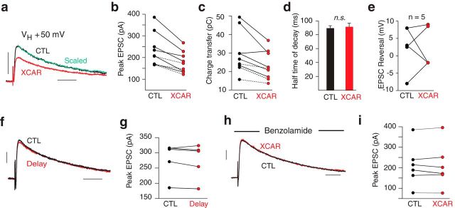
XCAR reduced NMDAR-mediated EPSCs. a, EPSC in XCAR (red) was reduced compared with control (CTL) at a VH of +50 mV. Scaled trace (green) shows no effect on time course. b, Effect of XCAR on peak EPSC in 9 cells. Dashed lines indicate use of human, recombinant type IV carbonic anhydrase in 3 cells. c, XCAR reduced charge transfer. d, Half-time of decay was unaffected by XCAR. e, XCAR had no effect on the EPSC reversal potential. f, Averaged EPSCs at 5–10 minutes (black) versus 17–22 minutes (delay, red) after breakthrough. g, Peak EPSC of 5 cells in delay experiments. h, XCAR had no effect on peak EPSC in presence of benzolamide. I, Peak EPSC in 6 cells exposed to XCAR in the presence of benzolamide. Calibrations: 100 pA/50 ms. VH = +50 mV for a–d and f–i.

