Figure 3.
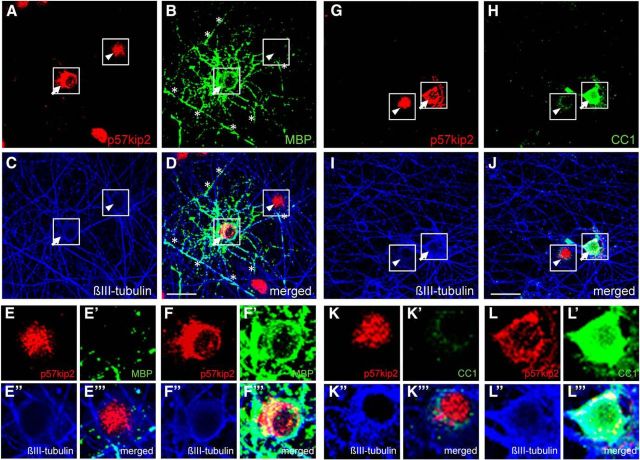
In vitro myelinating cocultures. A–J, Representative pictures of neuron–oligodendrocyte cocultures fixed and stained at DIV26, displaying active myelination of βIII-tubulin-positive axons (C,I). B, MBP-positive oligodendrocyte (arrow) and MBP-positive myelinated segments (asterisks). Myelin was formed by oligodendrocytes with cytoplasmic p57kip2 signals (arrow in D,F–F′′′), but not by cells with nuclear accumulation of p57kip2 (arrowhead in D,E–E′′′). G–J, Moreover, in this coculture p57kip2 expression could be confined to oligodendroglial cells and CC1-positive oligodendrocytes also exhibited cytoplasmic p57kip2 localization (J, arrow, L–L′′′). Scale bars, 30 μm.

