Figure 5.
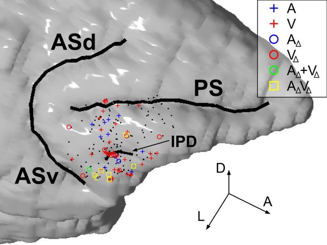
The locations of neurons that had significant activity in the audiovisual task are shown on a reconstruction of Monkey T's brain based on MRI. The neurons recorded from Monkey P were overlaid onto the reconstruction based on histologically confirmed anatomical landmarks. Color-coded dots are shown where activity was significantly modulated by the auditory component (A; A1 or A2) or the visual component (V; V1 or V2) of the vocalization movies (Eq. 1) and the auditory component change (AΔ; A1→A2 or A2→A1) or the visual component change (VΔ; V1→V2 or V2→V1) in Equation 4. AΔ + VΔ indicates significant main effects of both AΔ and VΔ, and AΔVΔ, a significant interaction. Black dots are neurons with no such effects. The three arrow lines indicate the directions of the anteroposterior (A), dorsoventral (D), and mediolateral (L) axes, respectively, and their lengths correspond to 5 mm each. PS, principal sulcus; ASd, dorsal arcuate sulcus; ASv, ventral arcuate sulcus; IPD, inferior prefrontal dimple.

