Fig. 3.
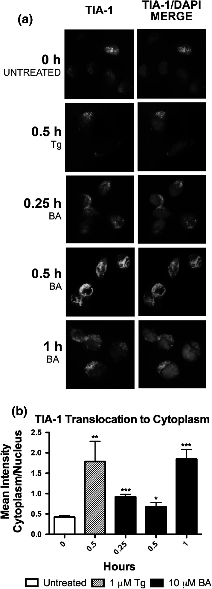
BA induces formation of TIA-1 positive stress granules. 10 µM BA induced formation of TIA-1 positive stress granules in DU-145 cells at 0.25, 0.5, and 1 h, indicated by TIA-1 protein (green) moving out of the nucleus (blue) with BA treatment. 1 µM thapsigargin (Tg) was used as a positive control, (n = 6–24 cells). All time points showed a significant increase. Pictures are representative of results. Significance differences were represented as *p < 0.05, **p < 0.01, and ***p < 0.001 and error bars represent standard deviations. (Color figure online)

