Abstract
As a finite-energy-bandgap alternative to graphene, semiconducting molybdenum disulfide (MoS2) has recently attracted extensive interest for energy and sensor applications. In particular for broad-spectral photodetectors, multilayer MoS2 is more appealing than its monolayer counterpart. However, little is understood regarding the physics underlying the photoresponse of multilayer MoS2. Here, we employ scanning photocurrent microscopy to identify the nature of photocurrent generated in multilayer MoS2 transistors. The generation and transport of photocurrent in multilayer MoS2 are found to differ from those in other low-dimensional materials that only contribute with either photovoltaic effect (PVE) or photothermoelectric effect (PTE). In multilayer MoS2, the PVE at the MoS2-metal interface dominates in the accumulation regime whereas the hot-carrier-assisted PTE prevails in the depletion regime. Besides, the anomalously large Seebeck coefficient observed in multilayer MoS2, which has also been reported by others, is caused by hot photo-excited carriers that are not in thermal equilibrium with the MoS2 lattice.
Two-dimensional materials have lately attracted tremendous attention owing to their unique structural and physical properties1. Understanding the basic physical phenomena as well as their intimate interplays in this new family of materials is essential for further exploitation of the materials towards electronic, photonic, and energy applications. Although it is largely graphene's tribute that has sparked the recent zeal in two-dimensional materials and their potential applications, the intrinsic shortcoming without a bandgap hampers its application in electronics2. For this reason, attention has been turned to other two-dimensional semiconducting transition metal dichalcogenides (TMDs) for their potential role in nanoelectronics, spintronics and optoelectronics3,4. Among them, molybdenum disulfide (MoS2), as a representative layered TMD, has a layer-dependent bandgap with a crossover from an indirect bandgap of 1.3 eV in its multilayer form to a direct bandgap of 1.8 eV when thinned to a monolayer5,6. Both single- and multi-layer MoS2 have been demonstrated to be suitable for electronic devices, such as field-effect transistors (FETs)7,8, photodetectors9,10, gas sensors11, etc. However, multilayer MoS2 is of greater potential than the monolayer counterpart from the fabrication viewpoint as well as the interesting physical properties. First, the electronic properties of multilayer MoS2 manifested by an indirect bandgap are relatively insensitive to layer thickness. Second, multilayer MoS2 is expected to carry higher drive current than monolayer MoS2 due to higher carrier mobility arising from interlayer screening effect12 and the 3-fold higher density of states at the conduction band minimum8,13.
With respect to optical properties, the subnanometer thickness of monolayer MoS2 yields a low absorption cross-section hence results in less photo-generated carriers, as compared to that of multilayer MoS2. Although fast photodetection has been achieved in monolayer-MoS2 transistors14, the photoresponsivity is expected to be lower than that of multilayer-MoS2 devices10, due to its direct bandgap and resultant shorter photocarrier recombination lifetime (~70 ps)15. For multilayer MoS2, intervalley scattering is predominant over direct electron-hole (e–h) recombination when incident light energy is larger than direct bandgap at K point, thus generating microsecond-order photocarrier lifetime of indirect recombination16. This was further proven by quenching of photoluminescence in multilayer MoS26. The long carrier lifetime would have a positive effect on photoresponsivity. There is therefore greater potential with multilayer MoS2 than monolayer MoS2 for applications in broad-spectral photodetectors.
Investigations of multilayer MoS2 and other TMD semiconductors are scarce and insufficient, compared to their monolayer counterparts, especially on optoelectronic properties. The origin of the observed photoresponse in MoS2 is still under debate. Most studies suggest that photocurrent generated in the MoS2 phototransistors results from photovoltaic effect (PVE)9,10,14,17,18, similarly to what has been found with other conventional semiconductors, e.g. Si, GaAs and GaN. Another photocurrent mechanism, i.e. photothermoelectric effect (PTE), is less discussed in the literature, in part due to the use of a broad-beam illumination experimental setup. Recent studies on graphene19,20 and carbon nanotubes21,22 indicate that PTE may play an important role in their photoresponse. As the amplitude of PTE relies on the temperature difference, multilayer MoS2 with a well-defined bulk, and therefore a reduced thermal sinking effect via the substrate, is expected to yield a higher temperature increase under illumination23. Because of the complex nature of MoS2, there is need to locally probe its photoresponse in order to allow the mechanisms of charge transport and collection in multilayer MoS2 to be unequivocally unveiled and, in particular, to discern PTE from PVE.
In order to elucidate the nature of photoresponse in thick MoS2 multilayers, we have employed scanning photocurrent microscopy (SPCM) in combination with ordinary current-voltage characterization to characterize the spatial dependence of photoresponse under various gate bias (Vg) conditions. At the metal-semiconductor interface, PVE is found to be predominant in the accumulation regime while thermal diffusion of photo-excited hot electrons is overwhelming and dictates the drifting current by the built-in electric field in the depletion regime.
Results
Device structure and characterization
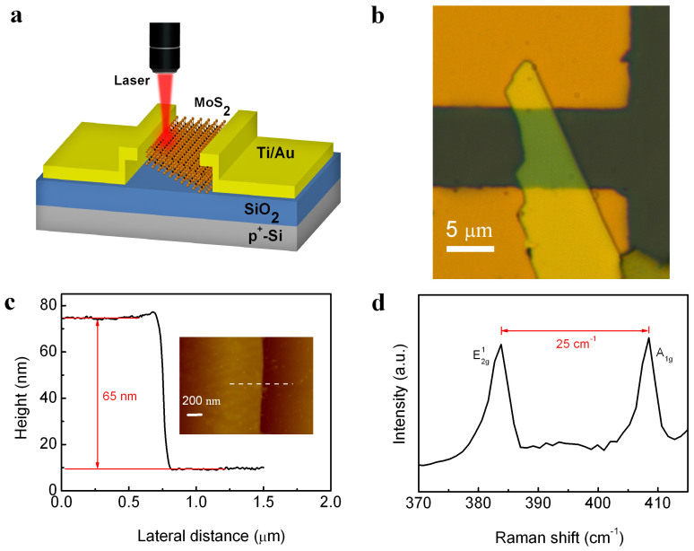
The schematic representation of a back-gate MoS2 transistor used in our work is shown in Fig. 1a while its optical microscope image is given in Fig. 1b. Isolated MoS2 flakes on a SiO2/Si substrate were exfoliated from a bulk MoS2 crystal using a conventional mechanical exfoliation technique24. The sample preparation and device fabrication are detailed in Methods. The thickness of the MoS2 flake in the device in Fig. 1b, as measured by atomic force microscopy (AFM), is approximately 65 nm (Fig. 1c) and its Raman spectrum in Fig. 1d shows two typical peaks (E2g1 and A1g) with a large separation of 25 cm−1, confirming that the multilayer nature of the MoS2 flake25.
Figure 1. Device structure and characterization.
(a) Schematic and (b) Optical image of the field-effect transistor based on multilayer MoS2. (c) AFM line scan along the dashed line across the boundary of the MoS2 flake in the inset. Inset: high-resolution AFM image of the MoS2 multilayer on SiO2/Si substrate. (d) Raman spectrum for the MoS2 multilayer on SiO2/Si substrate. The frequency separation between E12g and A1g peaks is 25 cm−1.
Photoelectrical stability of MoS2 transistor
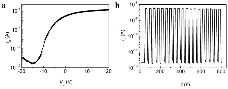
Electrical characterization of our MoS2 FETs was performed in ambient environment. Similar to other low-dimensional devices without surface protection26,27,28, MoS2 FETs are highly sensitive to extrinsic effects, especially adsorption of H2O/O2 from air29,30. These effects can induce current hysteresis in transfer characteristics and electrical stress instability of MoS2 FETs when measured in conventional DC mode (Supplementary Fig. S1). The electrical instability can lead to persistent photoconductivity when switching on and off the laser illumination and therefore, a reduction of photoresponsivity with switching time (Supplementary Figs. S3 and S4). Therefore, stability of the device under electrical stress is a prerequisite for practical optoelectronic applications of MoS2. Recently, electrical characterization with Vg pulses of alternating polarities i.e. AP mode, has been reported to distinctively reduce the device instability compared to using DC mode28,31,32. As shown in Supplementary Fig. S1 and Fig. 2a, the hysteresis of our MoS2 FETs is almost completely eliminated and stable electrical characteristics are obtained using the AP method. This method leads indeed to stable photoswitching behaviors when applying multiple illuminations (Fig. 2b and Supplementary Fig. S4), which can only be achieved in vacuum if MoS2 FETs are measured using the DC method33. Since the AP method is not based on any structural or chemical modification of the devices, as opposed to other measures employed to improve the device stability26,29,34, it is of great potential in leading to more repeatable and clearly interpretable photo-sensing signals. Therefore, all our photoelectrical measurements were performed in AP mode in order to avoid additional stressing of the devices.
Figure 2. Electrical characteristics and photoswitching behavior of MoS2 transistor.
(a) Transfer characteristics of the MoS2 transistor, measured at Vd = 1 V, with Vg sweeping in AP mode. (b) Stability of photoswitching behavior of the MoS2 transistor at Vd = 1 V and Vg = −15 V in AP mode. The laser spot was focused in the MoS2 channel with a laser power of 100 μW.
SPCM characterization of MoS2 transistor
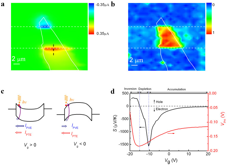
To identify the mechanism of photocurrent generation, SPCM was conducted at zero source-drain bias (Vd = 0 V) with a well-focused laser beam scanning over the device area. The short-circuit photocurrent and Raman spectra were recorded simultaneously as a function of laser position. To avoid possible photo-damage to the MoS2 during experiment35, the step size was set to be 1 μm. Figure 3a shows the photocurrent image measured at Vg = 15 V, where two photocurrent extrema of opposite polarities are observed at the electrode edges. As expected from conventional PVE, the opposite built-in electric fields from space charges at the source/drain contacts create an asymmetrical photocurrent characteristic. Such PVE-induced photocurrents localized near electrodes have been widely reported in devices based on silicon nanowires36 and carbon nanotubes37,38. However, it is remarkable to observe a significant photocurrent when the laser spot is centered inside the electrodes. The distance from the electrode edge is ~10 times larger than the laser spot size in order to make sure that the MoS2 channel is not illuminated. Besides, the light penetration depth is smaller than the thickness for the metal electrodes at the wavelengths used39. The absence of Raman signals from the MoS2 flake under the Ti/Au electrodes in Fig. 3b implies, therefore, that the observed photocurrent inside the electrodes could not arise from PVE at the MoS2-metal interface under the electrodes. Another photocurrent mechanism than PVE should be at work.
Figure 3. Photocurrent and Raman images of MoS2 transistor.
(a) Short-circuit scanning photocurrent images taken at Vg = 15 V with a laser power of 100 μW. (b) Raman mapping image with the normalized E12g peak intensity of the MoS2 multilayer. In panels (a) and (b), the dashed lines indicate the edges of the source and drain electrodes while the white lines mark the boundaries of MoS2. (c) Energy-band diagram of a multilayer- MoS2 transistor under light illumination in the accumulation (Vg > 0 V) and depletion (Vg < 0 V) regimes. Electron-hole pairs are generated in the space-charge region by light absorption and contribute to PVE and PTE photocurrents, which are indicated by blue and red arrows, respectively. (d) Gate-dependent Seebeck coefficient (S, black line) and the open-circuit photovoltage (VPH, red line) with a 100-μW laser illuminated at the contact edge. The vertical blue dotted lines separate the regimes of accumulation, depletion and inversion while the horizontal black dashed line indicates the border between the electron and hole conductions.
In fact, the absorption of laser photo energy by the metal electrodes in this manner could result in an inhomogeneous local temperature distribution. When a temperature gradient is generated by laser heating across a junction comprising two materials of different Seebeck coefficients, a thermal voltage driving a current through the device is established via thermoelectric effects. The polarity of thermoelectric current relies on the majority carriers (i.e. electrons or holes) and reverses sign in between the source and drain electrodes. As shown in Fig. 3a, the photocurrent image in the electrode area coincides with the optical surface topography of the MoS2 flake underneath the metal electrodes. This suggests that the PTE mechanism cannot be ignored in the photocurrent contribution. And the PTE-induced photocurrent is also expected to be intensified at the electrode edges where light can be absorbed by both materials and the heat dissipation is lower at the electrode edge than inside the metal electrodes.
The temperature rise at the MoS2-Ti/Au interface under laser illumination can be measured by Raman spectroscopy. Since the positions of the Raman-active phonons of MoS2 are sensitive to temperature40,41, the local temperature change induced by laser heating can be quantified. For calibration, the laser power was minimized in order to avoid heating effects in the Raman measurement and the temperature of the whole device was tuned by a temperature controller. The temperature-dependent and laser-power-dependent shifts of the Raman mode E2g1 are plotted in Supplementary Fig. S5. The linear temperature coefficient for mode E2g1 is extracted to be −0.015 cm−1/K in Supplementary Fig. S5c. In combination with the power-dependent coefficient of −0.74 cm−1/mW (Supplementary Fig. S5d), the temperature rise in the illuminated spot can be acquired. From this measurement, a 100-μW laser illumination at the MoS2-Ti/Au contact could result in a temperature increase by ~5 K. Thicker MoS2 layer could lead to a higher temperature rise due to the reduction of thermal coupling to the substrate23.
Photothermoelectric and photovoltaic effects
In the following discussion, we focus on two main competing mechanisms of photocurrent generation: PVE and PTE. The energy band diagrams of a MoS2 transistor with a Schottky barrier for various Vg conditions are schematically shown in Fig. 3c. For large positive Vg, the conduction band bends downward at the metal/semiconductor interface and the direction of the built-in electric field points from MoS2 to electrode. When Vg moves from positive to negative, the band bending direction changes from downward to upward and the built-in electric field reverses. The polarity of the photocurrent generated by PVE is dependent on the direction of the built-in electric field. However, in the PTE model, the generated photocurrent is equal to thermal voltage divided by device resistance R. Here, the thermal voltage is proportional to the local temperature increase in the junction, as well as to the difference in the Seebeck coefficients (S) between the MoS2 and the electrode20. Herein, S of the metal electrodes is negligible with respect to that of MoS242; the latter is gate-dependent and given by the Mott relation19,43 as follows:
 |
where, kB is the Boltzmann constant, q is elementary charge, T is absolute temperature, and EF is Fermi energy. The calculated S is plotted in Fig. 3d (black curve) using equation (1) in combination with the transfer characteristic curve of the device in dark. S shows a non-monotonic variation with gate bias, maximum at the threshold voltage, Vt (~−10 V) and reversing sign as the majority carrier changes from electron to hole when Vg < −15 V. At Vg > 0 V, both PVE and PTE contributions have the same sign, and therefore yield a large photocurrent, as illustrated in Fig. 3c. However, in the regime where the built-in electric field just switches the sign, the two contributions can have opposite signs and the photocurrent can then have either sign depending on their relative strengths.
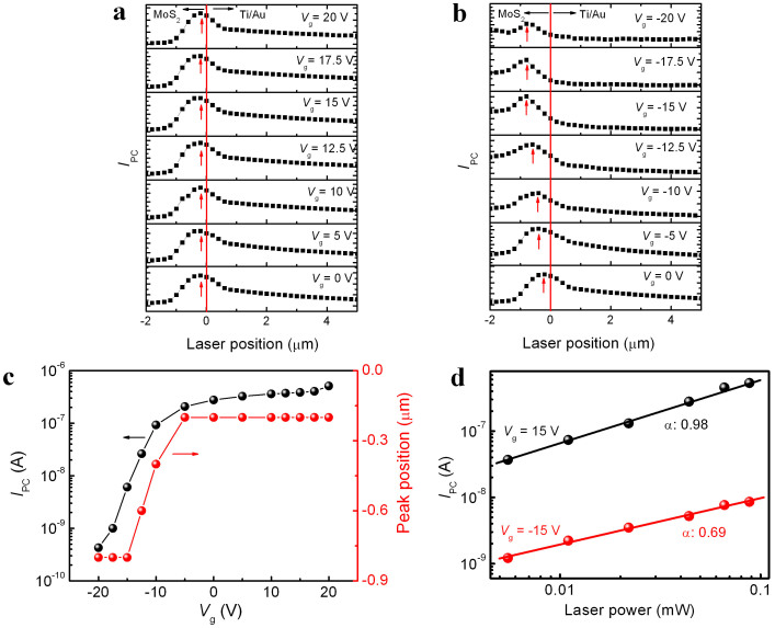
In order to gain more insights into the photocurrent generation mechanisms, we performed line-scan across the contact edge at a step size of 200 nm as shown by the black dashed line in Fig. 3a at various Vg (Figs. 4a–b). The peak position and peak amplitude of the photocurrents are extracted and plotted in Fig. 4c. The most striking feature in the plot is the movement of the peak position at the contact edge as Vg is decreased below −5 V. Concurrently, the peak amplitude decreases exponentially with Vg without sign flip. According to Fig. 3d, the thermoelectric photocurrent is expected to be negligible in the depletion regime (−15 V < Vg < −10 V) since S approaches zero in this region. As a result, PVE should dominate the photocurrent there. When Vg is changed from positive to negative, the built-in electric field reverses and the peak electric field at negative Vg moves away from the contact edge and extends more into the channel region compared to that at positive Vg (Supplementary Fig. S6). At Vg = −15 V, the photocurrent peak moves ~0.6 μm away from the contacts, which is in qualitative agreement with the movement of the simulated peak electric field. If the photocurrent generation is dominated by PVE in the depletion regime, the sign of photocurrent should reverse when (Vg − Vt) changes from positive to negative, which is however not observed experimentally. The contradicting polarity of the photocurrent suggests that the effect of the built-in field is less likely to be the dominant mechanism to determine the direction of the photocurrent in the depletion regime.
Figure 4. Gate-dependent photocurrent in MoS2 transistor.
(a) Photocurrent (IPC) line scans across the contact edge as indicated by the black dashed line in Fig. 3a at Vg from 0 V to 20 V and (b) Vg from 0 V to −20 V. The red arrows in panels (a) and (b) indicate the position of peak photocurrent. (c) Photocurrent peak position and amplitude in panels (a) and (b) varied with Vg. (d) The laser-power (P) dependency of the photocurrent with laser illuminated at the contact edge for Vg = 15 V and −15 V. The black and red lines are power-law fits with IPC ∝ Pα.
Thermoelectric power with two-carrier transport
Actually, the Mott formulism, which is established on the basis of thermal diffusion of one carrier type, is not valid for two types of carriers involved in the ambipolar transport. With light illumination, e–h pairs are generated when the photon energy exceeds the bandgap. Such excited carriers should contribute not only to the electrical conductivity, but also to the thermoelectric power through changing carrier concentration, which is more significant in the depletion regime. For such a two-carrier process, the thermoelectric power is given by44
 |
where, n and p are electron and hole concentrations, respectively, μn and μp are the corresponding mobilities, and Nc and Nv are the effective density of states in the conduction band and valence band, respectively, and An and Ap are thermoelectric constants of electrons and holes. When the concentration of one type of carriers is much higher than that of the opposite type of carriers, such as in the accumulation regime, S is then dominated by the majority carriers and equation (2) is reduced to the Mott relation43. If the electrons and the holes are equal in number, for example for photo-excited e–h pairs in the depletion regime, the effective carrier density of states would play a dominant role in determining the amplitude and sign of thermoelectric power. In multilayer MoS2, the minimum of the conduction band lies in the lower symmetry Q point in the k-space along the Γ-K line with a 6-fold valley degeneracy while the single valence band maximum is situated at the Γ point3. This results in an effective density of states in the conduction band almost 6 times that in the valence band by assuming an equal effective mass for electrons and holes. Therefore, PTE in the multilayer MoS2 is still dominated by the photo-excited electrons and both S and conductivity increase with illumination in the depletion regime. The unchanged polarity of photocurrent in the MoS2 FETs, when Vg is swept from positive to negative in Fig. 4b, reveals that PTE is predominant over PVE in the depletion regime. However, this photo-induced S in MoS2 is spatially inhomogeneous along the channel. In the space charge region, the built-in electric field reduces the e–h recombination rate, resulting in a higher S compared to that in the flat-band region. Recent studies22,45 reveal that a nonuniform S in the channel could produce an additional PTE photocurrent, which is different from that at the semiconductor-metal junction. This PTE photocurrent, as observed in Fig. 4b, moves with the gate bias in the depletion regime due to the photo-induced S depending on the electric field.
Hot photo-excited carrier inducing giant Seebeck coefficient
Apart from short-circuit photocurrent, open-circuit photovoltage can be a complementary diagnostic parameter to further probe the nature of the photoresponse. As shown in Fig. 3d (red curve), the gate-dependent photovoltage is measured at the contact edge. Identical to the photocurrent in Fig. 4c, the sign of photovoltage is not reversed during the gate sweeping and the maximum of photovoltage is shifted to more negative voltage (−15 V) compared to the gate-dependent S. Obviously, such a negative shift can be attributed to the photo-excited carriers. Since the main contribution of photovoltage arises from PTE in the depletion regime, the maximum S can be estimated from the photovoltage because there is only a constant temperature difference in between them. We have already determined that approximately a temperature increase by 5 K is obtained with a 100-μW laser illumination. The −0.18 V peak photovoltage would correspond to an S value of −3.6 × 10⁴ μV/K, which is one order of magnitude larger than the calculated maximum S value (black curve in Fig. 3d). This discrepancy in S has also been reported by Buscema et al.23. Nonetheless, the S value obtained for multilayer MoS2 is much larger than that of graphene19,20, carbon nanotubes21,22 and other thermoelectric materials46,47. In the estimation of S from the photovoltage data, the temperature increase induced by laser illumination is assumed to be equivalent to the temperature rise causing the Raman shift in the MoS2. However, this Raman-shift thermometry measures the phonon temperature instead of the electron temperature in the MoS2. It is the gradient in the electron, rather than phonon, temperature that ultimately drives the PTE effect. In the course of photo-excitation, the electrons and the phonons do not reach thermal equilibrium48. For example, in laser-illuminated graphene layers, electrons are much hotter than the surrounding crystal lattices and form a hot Fermion distribution19,49,50. Therefore, the measured phonon temperature is likely to underestimate the temperature of the electrons in the multilayer MoS2, resulting in a false anomalously giant S.
Strikingly, when Vg < −15 V, the calculated S in Fig. 3d reverses sign due to a weak hole inversion, while the polarity of photovoltage is opposite still staying positive. This suggests that the thermoelectric effect is still dominated by the excited hot electrons according to equation (2). In the strong electron accumulation regime (Vg > 0 V), the photovoltage saturates at a relative high value of −0.12 V, which results in a ratio of maximum-to- saturation photovoltage to be ~1.5. This is nearly two orders of magnitude smaller than that of calculated S (~102). This indicates that PVE rather than PTE is the dominant effect in the accumulation regime because the high electron concentration induced by gate voltage effectively cools the excited electrons due to the strong electron-electron interaction in reduced-dimension semiconductors51.
It has recently been demonstrated that the power-law relationship between photocurrent and incident laser power (P), i.e. IPC ∝ Pα, can be used as an alternative approach to characterizing the dominant mechanism for photocurrent generation52. The exponent α is unity for PVE and 2/3 for PTE. The laser-power dependency of the photocurrent at Vg = 15 V and −15 V is shown in Fig. 4d with the laser focused at the contact edge. The linear relationship at Vg = 15 V and the sublinear dependence on the laser power (α = 0.69) at Vg = −15 V further confirm that PVE dominates in the accumulation while PTE dictates in the depletion. This observation strikingly contradicts the common perception that one photocurrent mechanism (PVE or PTE) should dominate in the whole working regime, as is the case for nanotubes21,22,37,38 and graphene19,20. That PTE rather than PVE dominates in the depletion regime can be explained as follows. The optical absorption of MoS2 creates hot electrons. They can gain sufficient kinetic energy to overcome the potential barrier in the depletion regime. As a result, the hot electrons thermally diffusing along the channel generate the observed photocurrent in the depletion regime.
Discussion
A spatially resolved photoresponse of multilayer-MoS2 transistors has been investigated by using SPCM. When exposed to ambient environment, the transistors are electrically unstable causing undesired hysteresis and persistent photoconductivity when they are characterized using conventional DC measurement. Such instabilities are completely eliminated by using a novel pulsed-gate-bias method. As a result, a stable and repeatable photoresponse of MoS2 transistors is, for the first time, to our knowledge, achieved in ambient environment. This has allowed us to systematically explore the interplay of thermal, optical and electrical behaviors of multilayer MoS2. Both photovoltaic and photothermoelectric effects are found to be at work in the multilayer-MoS2 transistors, although they take dominant roles in different regimes of device operation. In the accumulation regime, the PVE at the MoS2-metal interface is the dominant mechanism whereas the hot-carrier-assisted PTE prevails in the depletion regime. This behavior is different from that observed in other low-dimensional materials that only contribute with either PVE or PTE. Moreover, the anomalously large Seebeck coefficient in MoS2, which has also been reported by others, is found to be the consequence of an oversimplification by assuming thermal equilibrium of the hot photo-excited carriers with the surrounding phonons. Our findings that shed light on the photoresponse mechanism of multilayer MoS2 may apply to other TMDs as well. They will also be helpful in advancing the art in light harvesting and photodetector applications.
Methods
Exfoliation and characterization of MoS2 thin flakes
Thin MoS2 flakes were peeled off from bulk MoS2 (SPI supplies) by mechanical exfoliation. They were subsequently transferred to a heavily doped p-type Si substrate with a 300-nm-thick thermally grown SiO2. The SiO2/Si substrate was pre-cleaned by sonication in acetone, isopropyl alcohol, and deionized water. The transferred MoS2 flakes were identified using an optical microscope (Keyence digital microscope VHX-600). The thickness of the MoS2 flakes was measured using AFM (Dimension 3100 with Nanoscope IIIa controller, Veeco) operated in tapping mode under ambient conditions. Raman spectra of MoS2 films were acquired by employing micro-Raman spectrometry (Renishaw inVia) in a backscattering configuration with a He-Ne 633 nm laser. The scattered signal was collected through a 100× objective and dispersed by a grating of 1800 lines/mm with a spectral resolution of ~1.0 cm−1 and a peak position accuracy of 0.1 cm−1. The power of the excitation laser line was kept well below 0.1 mW to avoid sample heating and an exposure time of 30 s was used to improve the signal-to-noise ratio.
Device fabrication and electrical characterization
In order to avoid the contamination from photolithography or electron-beam lithography, a 10-μm spacing copper grid was placed on top of the thin MoS2 flakes as a shadow mask for the electrode fabrication. A bilayer stack Ti/Au of 5/50 nm thickness was then deposited by means of electron-beam evaporator as the source and drain electrodes. Heavily doped Si substrate was used as the common back gate for the fabricated MoS2 FETs. Electrical characterization of the devices was carried out in a shielded probe station with Keithley 4200 semiconductor characterization system in the ambient environment.
SPCM characterization
Spatial dependence of photoresponse in the MoS2 transistors was obtained on the Renishaw Raman spectrometer equipped with the same electrical characterization system described above. A laser beam of 633-nm wavelength was focused to a diffraction-limited spot size of about 0.6 μm by using a long-working distance 50× objective lens. The incident laser power could be adjusted with the use of neutral density filters. A computer-controlled XYZ translational stage was used to acquire scanning photocurrent image and Raman mapping image simultaneously. When the laser spot was raster-scanned over the device area, the resultant current flow between electrodes and the Raman spectrum for each point in the scan were recorded simultaneously, thus generating spatially resolved images of photocurrent and Raman signal. All scanning photocurrent measurements were performed at room temperature in air.
Author Contributions
Z.-J.Q. conceived the research. Z.-J.Q. and Y.Z. designed the experiments. H.W., X.X. and Y.Z. carried out MoS2 device fabrication. Y.Z. performed AFM analysis, Raman measurement and SPCM characterizations. H.L. and L.W. performed TCAD simulation and first-principle calculations, respectively. Z.-J.Q. and Y.Z. analyzed the data. Z.-J.Q., S.-L.Z. and R.L. wrote the manuscript. All authors discussed the results and commented the manuscript.
Supplementary Material
Supplementary information
Acknowledgments
This work is supported partially by the Special Funds for Major State Basic Research Projects of China (No. 2011CBA00603), National Natural Science Foundation of China (Nos. 61204090 and 61171010), Shanghai Municipal Natural Science Foundation (No. 12ZR1402700), Fundamental Research Project of young teachers to enhance research capacity, Fudan University (No. 20520133248), and the Knut & Alice Wallenberg Foundation for a generous denotation to enable the “Swedish Graphene Initiative” project.
References
- Xu M., Liang T., Shi M. & Chen H. Graphene-like two-dimensional materials. Chem. Rev. 113, 3766–3798 (2013). [DOI] [PubMed] [Google Scholar]
- Schwierz F. Graphene transistors. Nature Nanotech. 5, 487–496 (2010). [DOI] [PubMed] [Google Scholar]
- Wang Q. H., Kalantar-Zadeh K., Kis A., Coleman J. N. & Strano M. S. Electronics and optoelectronics of two-dimensional transition metal dichalcogenides. Nature Nanotech. 7, 699–712 (2012). [DOI] [PubMed] [Google Scholar]
- Chhowalla M. et al. The chemistry of two-dimensional layered transition metal dichalcogenide nanosheets. Nature Chem. 5, 263–275 (2013). [DOI] [PubMed] [Google Scholar]
- Mak K. F., Lee C., Hone J., Shan J. & Heinz T. F. Atomically thin MoS2: a new direct-gap semiconductor. Phys. Rev. Lett. 105, 136805 (2010). [DOI] [PubMed] [Google Scholar]
- Splendiani A. et al. Emerging photoluminescence in monolayer MoS2. Nano Lett. 10, 1271–1275 (2010). [DOI] [PubMed] [Google Scholar]
- Radisavljevic B., Radenovic A., Brivio J., Giacometti V. & Kis A. Single-layer MoS2 transistors. Nature Nanotech. 6, 147–150 (2011). [DOI] [PubMed] [Google Scholar]
- Kim S. et al. High-mobility and low-power thin-film transistors based on multilayer MoS2 crystals. Nature Commun. 3, 1011 (2012). [DOI] [PubMed] [Google Scholar]
- Lopez-Sanchez O., Lembke D., Kayci M., Radenovic A. & Kis A. Ultrasensitive photodetectors based on monolayer MoS2. Nature Nanotech. 8, 497–501 (2013). [DOI] [PubMed] [Google Scholar]
- Choi W. et al. High-detectivity multilayer MoS2 phototransistors with spectral response from ultraviolet to infrared. Adv. Mater. 24, 5832–5836 (2012). [DOI] [PubMed] [Google Scholar]
- Li H. et al. Fabrication of single- and multilayer MoS2 film-based field-effect transistors for sensing NO at room temperature. Small 8, 63–67 (2012). [DOI] [PubMed] [Google Scholar]
- Castellanos-Gomez A. et al. Electric-field screening in atomically thin layers of MoS2: the role of interlayer coupling. Adv. Mater. 25, 899–903 (2013). [DOI] [PubMed] [Google Scholar]
- Kaasbjerg K., Thygesen K. S. & Jacobsen K. W. Phonon-limited mobility in n-type single-layer MoS2 from first principles. Phys. Rev. B 85, 115317 (2012). [Google Scholar]
- Yin Z. et al. Single-layer MoS2 phototransistors. ACS Nano 6, 74–80 (2012). [DOI] [PubMed] [Google Scholar]
- Korn T., Heydrich S., Hirmer M., Schmutzler J. & Schüller C. Low-temperature photocarrier dynamics in monolayer MoS2. Appl. Phys. Lett. 99, 102109 (2011). [Google Scholar]
- Shi H. et al. Exciton dynamics in suspended monolayer and few-layer MoS2 2D crystals. ACS Nano 7, 1072–1080 (2013). [DOI] [PubMed] [Google Scholar]
- Wu C.-C. et al. Elucidating the photoresponse of ultrathin MoS2 field-effect transistors by scanning photocurrent microscopy. J. Phys. Chem. Lett. 4, 2508–2513 (2013). [Google Scholar]
- Li H.-M. et al. Metal-semiconductor barrier modulation for high photoresponse in transition metal dichalcogenide field effect transistors. Sci. Rep. 4, 4041 (2014). [DOI] [PMC free article] [PubMed] [Google Scholar]
- Gabor N. M. et al. Hot carrier-assisted intrinsic photoresponse in graphene. Science 334, 648–652 (2011). [DOI] [PubMed] [Google Scholar]
- Xu X., Gabor N. M., Alden J. S., van der Zande A. M. & McEuen P. L. Photo-thermoelectric effect at a graphene interface junction. Nano Lett. 10, 562–566 (2010). [DOI] [PubMed] [Google Scholar]
- St-Antoine B. C., Ménard D. & Martel R. Position sensitive photothermoelectric effect in suspended single-walled carbon nanotube films. Nano Lett. 9, 3503–3508 (2009). [DOI] [PubMed] [Google Scholar]
- DeBorde T., Aspitarte L., Sharf T., Kevek J. W. & Minot E. D. Photothermoelectric effect in suspended semiconducting carbon nanotubes. ACS Nano 8, 216–221 (2014). [DOI] [PubMed] [Google Scholar]
- Buscema M. et al. Large and tunable photothermoelectric effect in single-layer MoS2. Nano Lett. 13, 358–363 (2013). [DOI] [PubMed] [Google Scholar]
- Novoselov K. S. et al. Electric field effect in atomically thin carbon films. Science 306, 666–669 (2004). [DOI] [PubMed] [Google Scholar]
- Li H. et al. From bulk to monolayer MoS2: evolution of Raman scattering. Adv. Funct. Mater. 22, 1385–1390 (2012). [Google Scholar]
- Kim W. et al. Hysteresis caused by water molecules in carbon nanotube field-effect transistors. Nano Lett. 3, 193–198 (2003). [Google Scholar]
- Levesque P. L. et al. Probing charge transfer at surfaces using graphene transistors. Nano Lett. 11, 132–137 (2011). [DOI] [PubMed] [Google Scholar]
- Qu M., Li H., Liu R., Zhang S.-L. & Qiu Z.-J. Interaction of bipolaron with the H2O/O2 redox couple causes current hysteresis in organic thin-film transistors. Nature Commun. 5, 3185 (2014). [DOI] [PubMed] [Google Scholar]
- Late D. J., Liu B., Ramakrishna Matte H. S. S., Dravid V. P. & Rao C. N. R. Hysteresis in single-layer MoS2 field effect transistors. ACS Nano 6, 5635–5641 (2012). [DOI] [PubMed] [Google Scholar]
- Cho K. et al. Electric stress-induced threshold voltage instability of multilayer MoS2 field effect transistors. ACS Nano 7, 7751–7758 (2013). [DOI] [PubMed] [Google Scholar]
- Mattmann M. et al. Pulsed gate sweep strategies for hysteresis reduction in carbon nanotube transistors for low concentration NO2 gas detection. Nanotechnology 21, 185501 (2010). [DOI] [PubMed] [Google Scholar]
- Liu Z., Qiu Z.-J., Zhang Z.-B., Zheng L.-R. & Zhang S.-L. Mobility extraction for nanotube TFTs. IEEE Elec. Dev. Lett. 32, 913–915 (2011). [Google Scholar]
- Zhang W. et al. High-gain phototransistors based on a CVD MoS2 monolayer. Adv. Mater. 25, 3456–3461 (2013). [DOI] [PubMed] [Google Scholar]
- Radisavljevic B. & Kis A. Mobility engineering and metal-insulator transition in monolayer MoS2. Nature Mater. 12, 815–820 (2013). [DOI] [PubMed] [Google Scholar]
- Castellanos-Gomez A. et al. Laser-thinning of MoS2: on demand generation of a single-layer semiconductor. Nano Lett. 12, 3187–3192 (2012). [DOI] [PubMed] [Google Scholar]
- Ahn Y., Dunning J. & Park J. Scanning photocurrent imaging and electronic band studies in silicon nanowire field effect transistors. Nano Lett. 5, 1367–1370 (2005). [DOI] [PubMed] [Google Scholar]
- Freitag M. et al. Imaging of the Schottky barriers and charge depletion in carbon nanotube transistors. Nano Lett. 7, 2037–2042 (2007). [DOI] [PubMed] [Google Scholar]
- Balasubramanian K., Burghard M., Kern K., Scolari M. & Mews A. Photocurrent imaging of charge transport barriers in carbon nanotube devices. Nano Lett. 5, 507–510 (2005). [DOI] [PubMed] [Google Scholar]
- Sanders A. W. Optical properties of metallic nanostructures. Ph.D. thesis, Yale University, New Haven, Connecticut, 2007.
- Yan R. et al. Thermal conductivity of monolayer molybdenum disulfide obtained from temperature-dependent Raman spectroscopy. ACS Nano 8, 986–993 (2014). [DOI] [PubMed] [Google Scholar]
- Sahoo S., Gaur A. P. S., Ahmadi M., Guinel M. J.-F. & Katiyar R. S. Temperature-dependent Raman studies and thermal conductivity of few-layer MoS2. J. Phys. Chem. C 117, 9042–9047 (2013). [Google Scholar]
- Cusack N. & Kendall P. The absolute scale of thermoelectric power at high temperature. Proc. Phys. Soc. 72, 898–901 (1958). [Google Scholar]
- Ashcroft N. W. & Mermin N. D. Solid State Physics Ch. 13 (Thomson Learning Inc., London, 1976). [Google Scholar]
- Harper J. G., Matthews H. E. & Bube R. H. Photothermoelectric effects in semiconductors: n- and p-type silicon. J. Appl. Phys. 41, 765–770 (1970). [Google Scholar]
- Nanot S. et al. Broadband, polarization-sensitive photodetector based on optically-thick films of macroscopically long, dense, and aligned carbon nanotubes. Sci. Rep. 3, 1335 (2013). [DOI] [PMC free article] [PubMed] [Google Scholar]
- Ohta H. et al. Giant thermoelectric seebeck coefficient of a two-dimensional electron gas in SrTiO3. Nature Mater. 6, 129–134 (2007). [DOI] [PubMed] [Google Scholar]
- Pernstich K. P., Rössner B. & Batlogg B. Field-effect-modulated Seebeck coefficient in organic semiconductors. Nature Mater. 7, 321–325 (2008). [DOI] [PubMed] [Google Scholar]
- Eesley G. L. Generation of nonequilibrium electron and lattice temperatures in copper by picosecond laser pulses. Phys. Rev. B 33, 2144–2151 (1986). [DOI] [PubMed] [Google Scholar]
- Sun D. et al. Ultrafast hot-carrier-dominated photocurrent in graphene. Nature Nanotech. 7, 114–118 (2012). [DOI] [PubMed] [Google Scholar]
- Song J. C. W., Rudner M. S., Marcus C. M. & Levitov L. S. Hot carrier transport and photocurrent response in graphene. Nano Lett. 11, 4688–4692 (2011). [DOI] [PubMed] [Google Scholar]
- Liang Y., Huang S., Soklaski R. & Yang L. Quasiparticle band-edge energy and band offsets of monolayer of molybdenum and tungsten chalcogenides. Appl. Phys. Lett. 103, 042106 (2013). [Google Scholar]
- Patil V., Capone A., Strauf S. & Yang E.-H. Improved photoresponse with enhanced photoelectric contribution in fully suspended graphene photodetectors. Sci. Rep. 3, 279 (2013). [DOI] [PMC free article] [PubMed] [Google Scholar]
Associated Data
This section collects any data citations, data availability statements, or supplementary materials included in this article.
Supplementary Materials
Supplementary information


