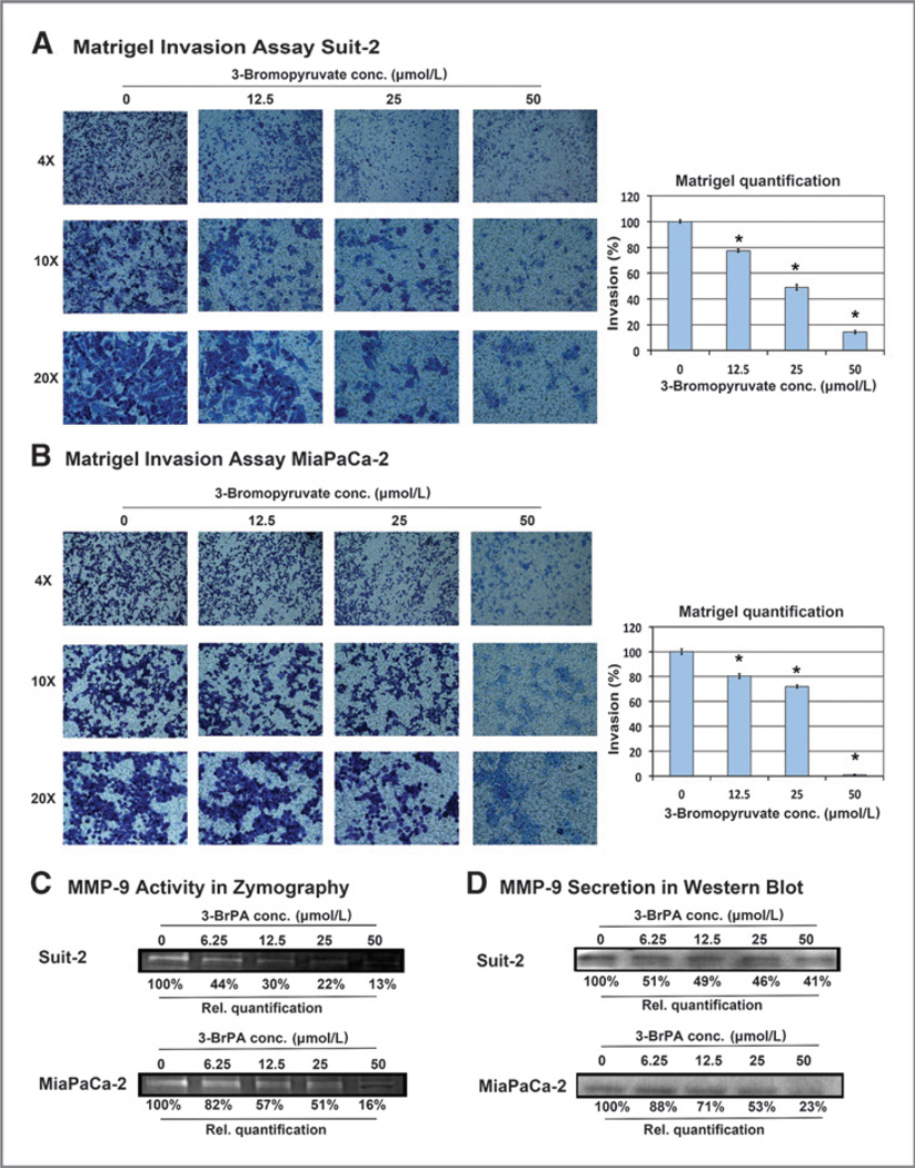
Figure 3.
Effects of 3-BrPA on cell invasiveness. MiaPaCa-2 (A) and Suit-2 (B) cells were plated into a Boyden invasion chamber. Incubation overnight was followed by treatment with 3-BrPA for 48 hours (MiaPaCa-2) or 72 hours (Suit-2). Invaded cells on the bottom side of the membrane of the invasion insert were stained using a Giemsa-like staining. Images show invaded cells at 4×, 10×, and 20× magnification. Relative quantification of invasion was calculated by measuring the area of stained cells in the entire field of view at 10×. MMP-9 activity and secretion were determined in the concentrated supernatant of MiaPaCa-2 and Suit-2 cells by zymography (C) and Western blot analysis (D). *, indicates statistically significant differences (P < 0.05).

