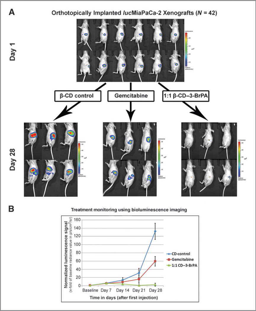
Figure 4.
In vivo efficacy of β-CD–3-BrPA. A total of 42 male nude mice were orthotopically implanted with a total of 1.5 × 10⁶ lucMiaPaCa-2 cells. After one week of xenograft growth, tumors were confirmed using bioluminescence imaging (BLI). A representative number of animals are shown in (A). Animals were randomized to receive β-CD–3-BrPA (n = 21), free 3-BrPA (n = 7), gemcitabine (n = 7), and β-CD (n = 7). Animals were imaged once per week over the course of 28 days. The overall progress of the signal is demonstrated in B. Animals treated with free 3-BrPA showed high treatment-related toxicity and did not survive in statistically relevant numbers to be included in the final image analysis.

