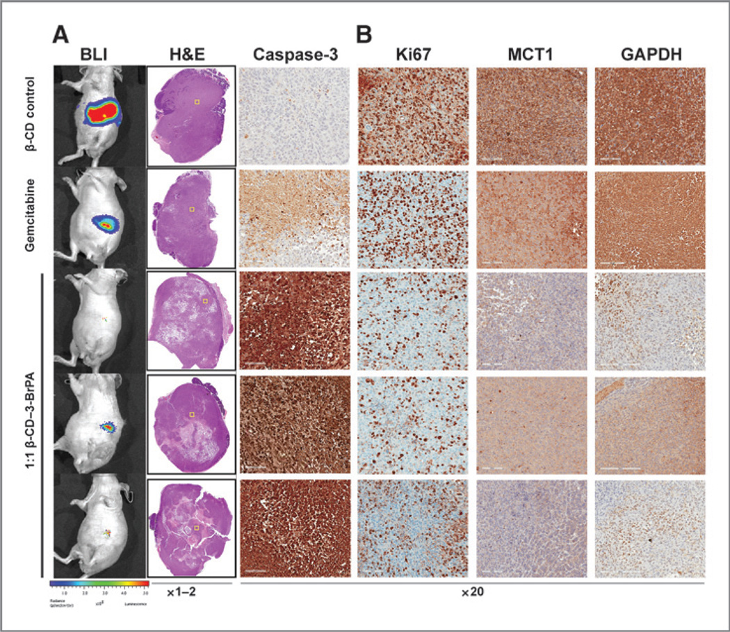
Figure 5.
Ex vivo pathologic and immunohistochemical tumor analysis. The H&E staining of tumors treated with β-CD, gemcitabine or β-CD–3-BrPA (3 representative tumors are shown here under A) demonstrated the treatment effects of β-CD–3-BrPA. The yellow squares within the H&E-stained whole-tumor overviews indicate the areas magnified for further analysis of the antitumoral effects of the drugs, which was confirmed by the staining for cleaved caspase-3 and Ki-67 (B). In addition, the marked reduction of GAPDH as the primary target of 3-BrPA as well as MCT-1 as the specific transporter becomes apparent (B). Of note, only areas that did not show complete/liquefied necrosis were selected for analysis in higher magnification.

