FIG 5.
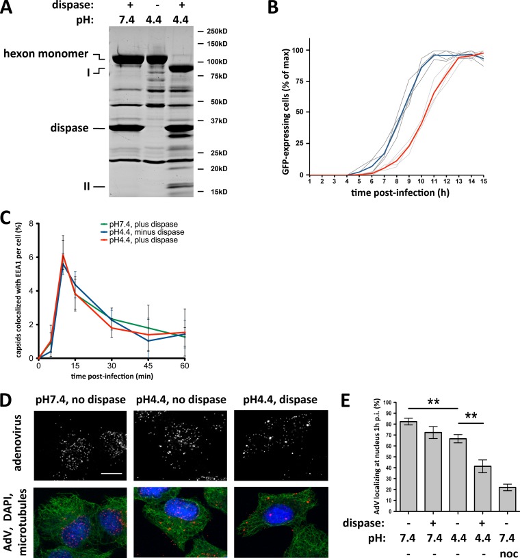
Dispase treatment reduces capsid redistribution to the nucleus. (A) pH-dependent dispase cleavage of AdV5 capsid hexon. Purified AdV5 capsids were treated with dispase at pH 7.4 or pH 4.4 or kept at pH 4.4 alone before being boiled in PSB and subjected to SDS-PAGE and Coomassie blue staining to show total capsid proteins. Hexon was hydrolyzed by dispase only at pH 4.4 (85-kDa and 15-kDa products, marked with I and II, respectively), whereas no proteolysis was detectable at pH 7.4 or pH 4.4 without the protease. (B) Effect of dispase cleavage on infectivity. AdV5-GFP capsids were treated with dispase at pH 7.4 or pH 4.4 and used to infect A549 cells, which were continuously imaged for up to 15 h p.i. Graphs represent relative counts of GFP-expressing cells. Dispase-cleaved capsids (light gray lines; the red line is the average) induce GFP expression ∼2 h later than control capsids (dark gray lines; the blue line is the average). (C) Normal cell entry by dispase-treated capsids. A549 cells were infected with untreated or dispase-treated AdV5 capsids, fixed at the indicated times p.i., and processed for immunofluorescence microscopy using antihexon and anti-EEA1 (early endosome antigen 1) antibodies. The graph shows the percentage of colocalization of AdV capsids with EEA1 over time postinfection and indicates similar profiles independent of dispase treatment. (D) Effect of dispase treatment on capsid redistribution to nucleus. A549 cells were infected with untreated or dispase-treated AdV5 capsids, fixed at 60 min p.i., and processed for immunofluorescence microscopy using antihexon antibody to localize capsids within the cells. Cells were also stained for microtubules (antitubulin) and DNA (DAPI). Dispase-treated capsids remained dispersed, whereas control capsids redistributed to the nucleus. Bar, 10 μm. (E) Quantification of virus redistribution. The nuclear localization of AdV5 particles was moderately reduced by pretreatment with dispase at pH 7.4 or without dispase at pH 4.4. More substantial inhibition of virus redistribution resulted from treatment of virus with dispase at pH 4.4 compared to the untreated pH 4.4 control (n = 3; means ± standard deviations). The nocodazole control used to depolymerize microtubules showed near-complete inhibition of virus redistribution to the nucleus. Asterisks mark P values of <0.01.

