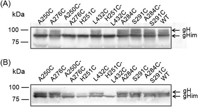
FIG 3.
Western blot analyses. Lysates of RK13 cells transfected with wild-type (WT) or mutated gH expression plasmids (A) or cotransfected with gH and gL expression plasmid (B) were separated by SDS-PAGE. Gels were incubated with a gH-specific rabbit antiserum. The signals of immature (gHim) and mature gH are labeled by arrows, and molecular masses of marker proteins are indicated.

