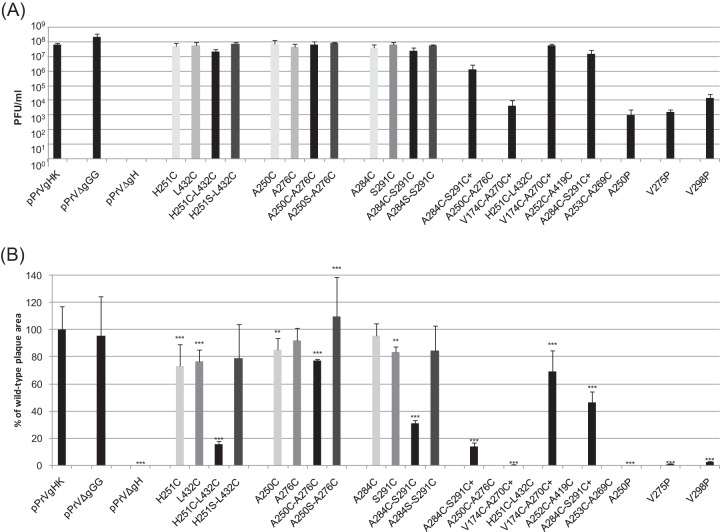
FIG 7.
Final titers and plaque sizes of PrV mutants. (A) RK13 cells were infected with pPrV-gHK, pPrV-ΔgGG, pPrV-ΔgHABF, or virus recombinants containing the indicated gH mutations. Progeny virus titers were determined 48 h after infection at an MOI of 0.1 by plaque assays on RK13 or RK13-gH/gL cells. Shown are the mean results of three experiments and standard deviations. (B) Areas of 30 plaques or foci of infected RK13 cells per virus were determined 48 h after infection by fluorescence microscopy, and mean sizes were plotted as percentages of wild-type (pPrV-gHK) sizes. Standard deviations (vertical lines) and significant size reductions (*, P < 0.05; **, P < 0.01; ***, P < 0.001) are indicated.

