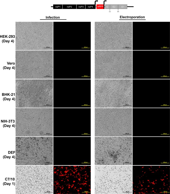
FIG 7.
Infection of vertebrate and mosquito cell lines with EILV-eRFP with SINV structural ORF. Each cell line was infected at an MOI of 10 or electroporated with 10 μg of genomic RNA. Phase-contrast (left) and fluorescence (right) micrographs were taken at 1 dpi (C7/10 cells) or 4 dpi (vertebrate cells).

