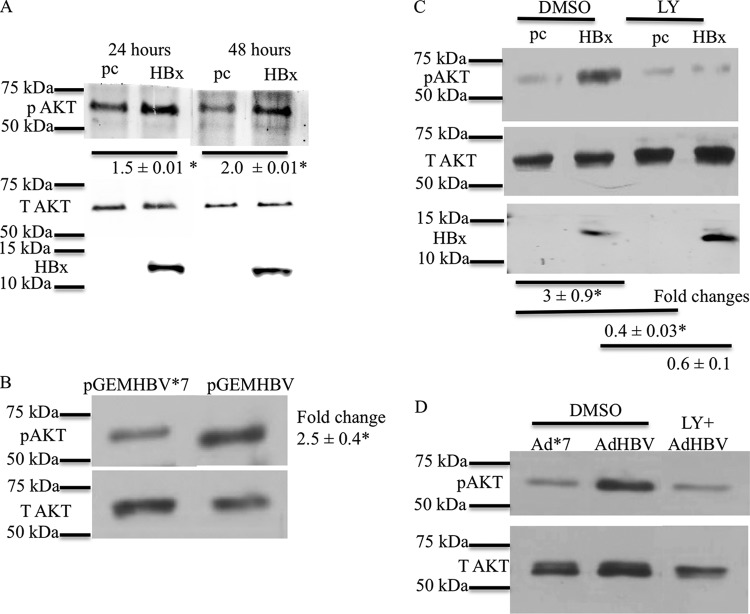
FIG 1.
HBx activates AKT in primary rat hepatocytes. (A) Primary rat hepatocytes were transfected with the HBx expression plasmid or the pcDNA control vector. The cells were collected at different time points, and the levels of pAKT (Ser473) were measured by Western blot analyses. T AKT, total AKT. (B) Primary rat hepatocytes were transfected with the pGEMHBV or pGEMHBV*7 (HBx-deficient HBV) expression plasmid. Western blot analysis was performed to check the levels of pAKT in pGEMHBV- and pGEMHBV*7-transfected cells. (C) Primary rat hepatocytes were transfected with the HBx expression plasmid or the pcDNA control vector. The cells were treated with either the DMSO control or LY294002 (LY) (PI3K inhibitor), and Western blot analysis was performed to check the levels of pAKT in HBx- and pcDNA-transfected cells. (D) Primary human hepatocytes were infected with AdHBV or Ad*7 (without HBx) and then treated with either the vehicle control or LY294002. Western blot analysis was conducted to assess the levels of AKT activation. Each Western blot shows one representative result from at least two independent experiments. The band intensities in each Western blot were measured by using ImageJ software; the band intensities of pAKT were divided by the band intensities of total AKT, and the ratios are represented in the terms of fold differences. The differences indicated are average fold changes from three independent experiments ± standard errors. An asterisk represents a P value of <0.05, determined by Student's t test.

