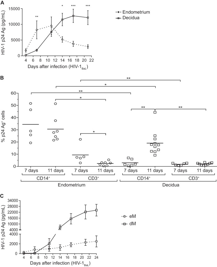
FIG 1.
Infection of endometrial and decidual mononuclear cells and macrophages. (A and B) Endometrial and decidual mononuclear cells were exposed to HIV-1BaL at an MOI of 10−3. (A) The HIV-1 p24 Ag was measured by ELISA in the culture supernatants of endometrial (n = 11) and decidual (n = 16) mononuclear cells over time. The mean and the standard error of the mean of the viral production are displayed. (B) Intracellular staining of the p24 Ag was performed on days 7 and 11 postinfection in endometrial and decidual mononuclear cells. The percentage of p24 Ag+ cells among CD14+ cells or CD3+ cells is shown (n = 5 on day 7 and n = 7 on day 11 for the endometrium; n = 6 on day 7 and n = 11 on day 11 for the decidua). Each circle represents one endometrial sample and each square one decidual sample. The means are displayed. *, P < 0.05; **, P < 0.005; ***, P < 0.0005. (C) Endometrial (eM, n = 4) and decidual (dM, n = 17) purified macrophages were exposed to HIV-1BaL at an MOI of 10−3. The HIV-1 p24 Ag was measured by ELISA in the culture supernatants over time.

