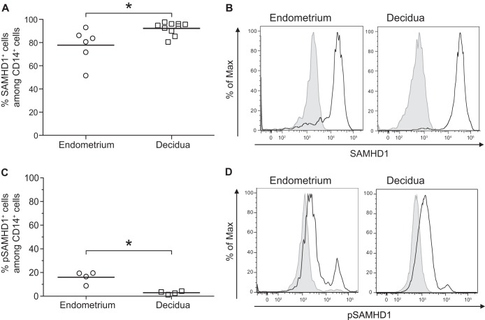
FIG 5.
Expression of SAMHD1 in decidual and endometrial macrophages. The expression of SAMHD1 and the phosphorylated form of SAMHD1 at residue Thr592 (pSAMHD1) was analyzed on CD45+ CD14+ cells by flow cytometry staining of total endometrial or decidual cells. An isotype-matched IgG control was used to confirm the specificity of the anti-SAMHD1 and the anti-pSAMHD1 Thr592. The mean percentages of SAMHD1 (A) and pSAMHD1 (C) expression among the CD14+ cells are displayed. Each circle represents one endometrial sample (n = 6 for SAMHD1 and n = 4 for pSAMHD1) and each square one decidual sample (n = 10 for SAMHD1 and n = 4 for pSAMHD1). *, P < 0.05. (B and D) A representative histogram is shown for one endometrial sample and for one decidual sample. The black line represents SAMHD1 (B) or pSAMHD1 (D) expression and shaded gray the isotype-matched control IgG.

