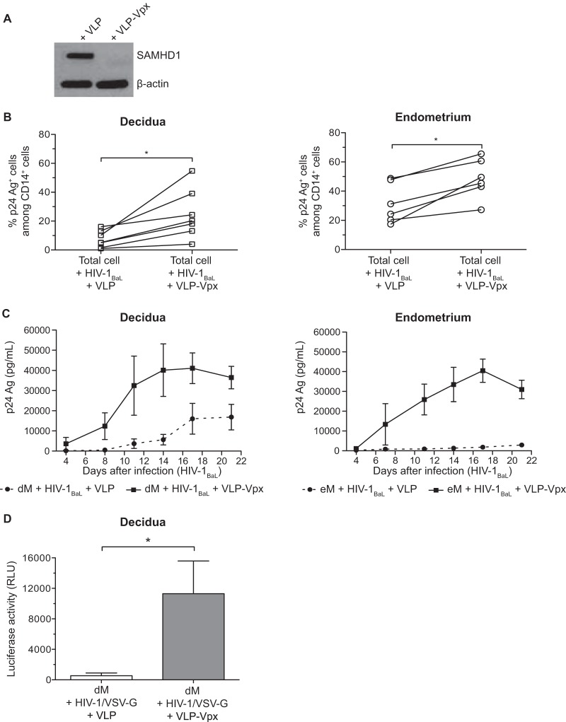
FIG 6.
SAMHD1 degradation and infection of decidual and endometrial cells in the presence of VLP-Vpx. (A) Control VLP or VLP-Vpx was added to purified decidual CD14+ cells, and after 72 h, SAMHD1 expression was analyzed by Western blotting. (B) Decidual and endometrial cells were exposed to HIV-1BaL at an MOI of 10−3 in the presence of control VLP or VLP-Vpx. Intracellular staining of the p24 Ag was performed on day 11 postinfection. The percentage of p24 Ag+ cells among CD14+ cells of each sample (n = 6 for the endometrium and n = 7 for the decidua) is displayed. (C) Purified eM and dM were exposed to HIV-1BaL at an MOI of 10−3 in the presence of control VLP (dashed line) or VLP-Vpx (full line). Values represent the means of the p24 Ag concentrations over time (n = 3 for the endometrium and n = 4 for the decidua). (D) Purified dM were exposed to the HIV-1/VSV-G pseudotype (600 ng of p24 Ag/106 cells) in the presence of control VLP or VLP-Vpx. The mean luciferase activity is given for 6 samples, in relative light units (RLU). Bars indicate standard errors of the means. *, P < 0.05.

