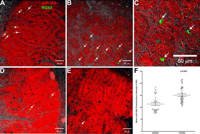
FIG 4.
Localization of miR-99b to both duodenal epithelium and lamina propria cells (A, B, D, and E) in both VEH/SIV (FE07, HT49) and THC/SIV (HN56, HV47) macaques. Note the increased miR-99b staining intensity in the THC/SIV macaques (D and E) compared to the VEH/SIV macaques (A and B). Increased numbers of duodenal crypt epithelial cells from both VEH/SIV macaques stained positive for NOX4 (A and B). (C) A close-up view of the lower right quadrant of panel B clearly shows NOX4 staining localized to the epithelial cell cytoplasm. In contrast, duodenal crypt epithelial cells from both THC/SIV macaques (D and E) show occasional NOX4+ cells. Small white arrows (A, B, D, and E) point to NOX4+ crypt epithelial cells. Epithelial NOX4+ (A to C, D, and E) staining cells are shown in green (Alexa Fluor-488). Epithelial miR-99b+ (A to C, D, and E) staining cells are shown in red (permanent red substrate). All panels are shown at a magnification of ×40. (F) Quantitation of cells and regions of interest (ROI), which were labeled by an LNA-modified miR-99b probe using Volocity 5.5 software, revealed significantly elevated miR-99b expression in THC/SIV compared to VEH/SIV macaques. Several ROI were hand drawn on the epithelial and LPL regions in the images from duodenum. Image analysis data were analyzed using nonparametric Wilcoxon's rank sum test.

