
Figure 5.
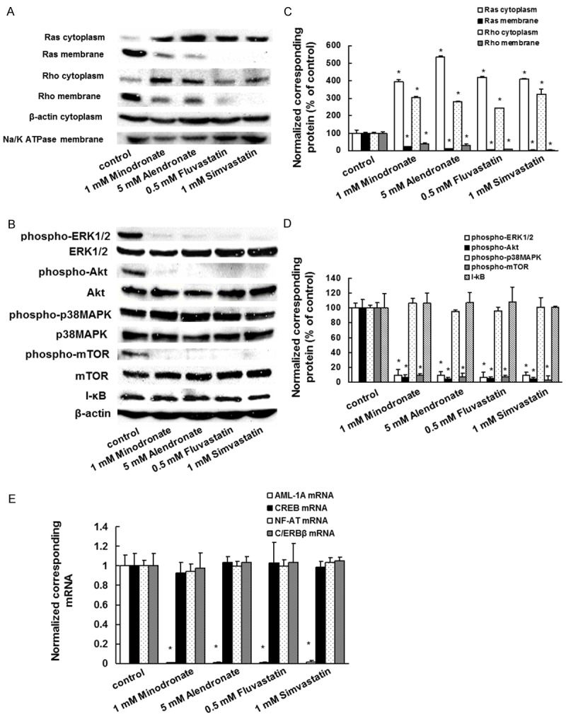
Effect of bisphosphonates and statins on Ras/Rho membrane localization, activation of signaling molecules, and expression of AML-1A mRNA in IM9 cells. The cells were incubated with 1 μM minodronate, 5 μM alendronate, 0.5 μM fluvastatin, or 1 μM simvastatin for 3 days. A. The cytoplasmic fractions and membrane fractions were extracted and then subjected to SDS-PAGE/immunoblotting with anti-Ras and anti-Rho antibodies. Anti-Na/K-ATPase antibody or Anti-β-actin antibody was used as the membrane or cytoplasm internal standard, as the primary antibody to detect Na/K-ATPase or β-actin protein. B. The cytoplasmic fractions were extracted and then subjected to SDS-PAGE/immunoblotting with antibodies against phosphorylated ERK1/2 (phospho-ERK1/2), phosphorylated Akt (phospho-Akt), phosphorylated p38MAPK (phospho-p38MAPK), I-κB, phosphorylated mTOR (phospho-mTOR), ERK, Akt, p38MAPK, mTOR, and β-actin. C. Quantification of the amount of Ras or Rho, normalized to the amounts of the corresponding proteins, respectively. The results are representative of 5 independent experiments. *P < 0.01, as compared to controls (ANOVA with Dunnett’s test). D. Quantification of the amount of phospho-ERK1/2, phospho-Akt, phospho-p38MAPK, phospho-mTOR, or I-κB, normalized to the amounts of the corresponding proteins, respectively. The results are representative of 5 independent experiments. *P < 0.01, as compared to controls (ANOVA with Dunnett’s test). E. A total RNA was extracted, and AML-1A, CREB, NF-AT, and C/ERBβ mRNA levels were determined by real-time PCR. IM-9 cells treated with minodronate, alendronate, fluvastatin, or simvastatin for 3 days. The results are representative of 5 independent experiments. *P < 0.01, as compared to controls (ANOVA with Dunnett’s test).