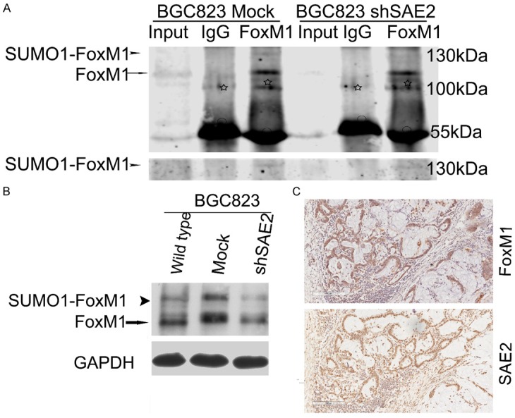
Figure 6.

FoxM1 is SUMOylated in vivo, and SAE2 was positively correlated with SUMO1-FoxM1, FoxM1 expression levels in GC. A. Cell lysates were immunoprecipitated and subsequently immunoblotted with the indicated antibodies. SUMOylated FoxM1 and non-SUMOylated FoxM1 are indicated by an arrowhead and an arrow, respectively. White asterisks and circles indicate nonspecific blot and IgG heavy chain, respectively. B. Western blotting analysis of SUMOylated-FoxM1 (SUMO1-FoxM1), total FoxM1 proteins in BGC823. C. One representative case of FoxM1 and SAE2 staining in serial sections of surgically resected GC tissue.
