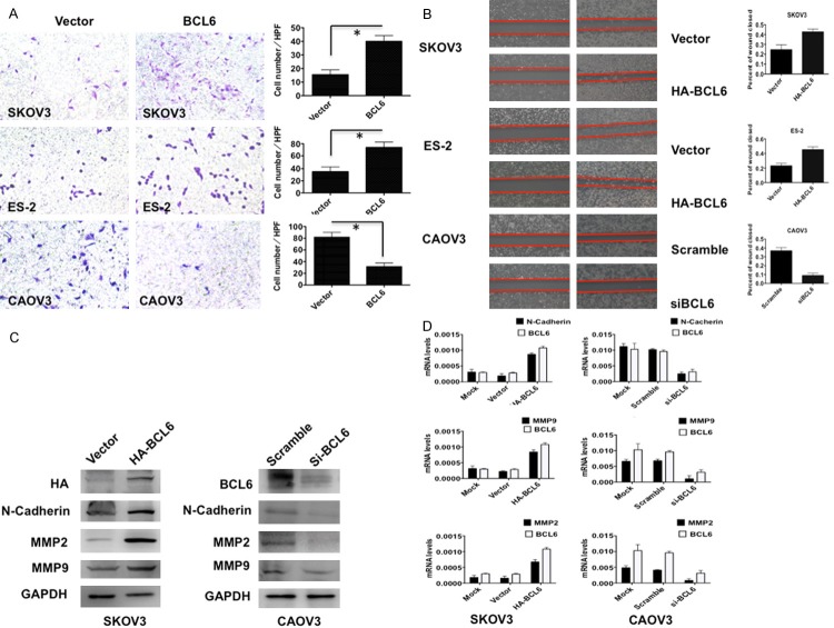
Figure 5.
BCL6 induced tumor cell invasion and migration in ovarian carcinoma. A: Representative images (left) and quantification (right) of transwell invasion assays for indicated cells. (Scale bars = 50 μm). *p < 0.01. B: Representative images (left) and quantification (right) of wound-healing assays for indicated cells. (Scale bars = 400 μm). *p < 0.01. C: The immunoblotting results of N-Cadherin, MMP2 and MMP9 in indicated cells. GAPDH was used as reference. D: The mRNA levels of N-Cadherin, MMP2 and MMP9 in indicated cells. GAPDH was used as reference.

