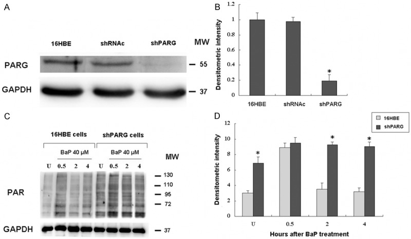
Figure 1.

Silencing of poly(ADP-ribose) glycohydrolase (PARG) by shRNA in 16HBE cells. 16HBE cells were transfected with shRNA oligos for PARG as previously reported [14]. A: Western blotting analysis of shRNAc (lane 1) and shPARG (lane 2) cell lysates probed with antibodies against PARG and GAPDH. B: Densitometric quantification of PARG protein bands from A. C: The 16HBE cells and shPARG cells were treated with 40 µmol/L BaP for 20 min, then analyzed for levels of PAR by immunoblot from 0.5-4 h. Equal protein loading per lane was verified by immunoblotting detection of GAPDH. D: Densitometric quantification of PAR levels from C. *indicates a significant difference (P < 0.05) between 16HBE and shPARG cells. Error bars represent the SEM. All experiments were repeated at least 3 times with similar results.
