
Figure 5.
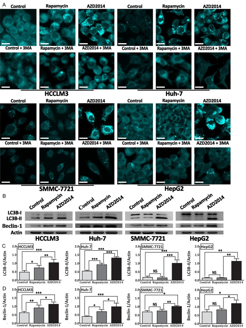
AZD2014 induces autophagy in HCC cells. (A) MDC-labeled vacuoles were induced by AZD2014 and inhibited by autophagy inhibitor (3-MA). HCCLM3, Huh-7, SMMC-7721, and HepG2 cells were treated with AZD2014 or rapamycin at concentrations of 100, 440, 140 and 600 nM, respectively, for 48 hours in the presence or absence of 3-MA, and then stained with MDC. Cells were immediately observed under a confocal microscope. Cells in the control group were treated with DMSO. bars, 20 μm. (B) Expression of LC3B (LC3B-I and LC3B-II) and Beclin-1 determined by immunoblot analysis in HCC cells treated as described above. ImageJ densitometric analysis was used for the measurement of LC3B-II/Actin (C), and Beclin-1/Actin (D) ratios from immunoblots. Each column was from the mean of at least three independent experiments; bars, SD. *P < 0.05, **P < 0.01, ***P < 0.001, NS = not significant.