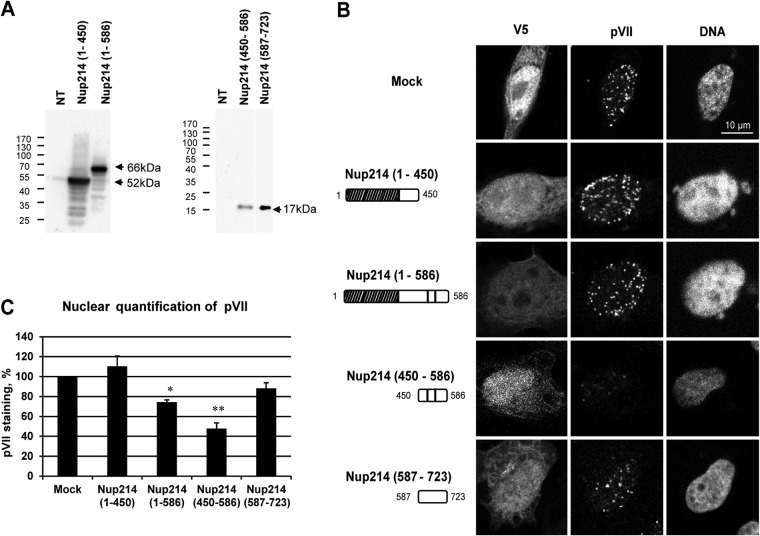
FIG 6.
Reduction of nuclear localization of protein VII in HeLa cells overexpressing soluble Nup214 proteins. HeLa cells were transfected with expression plasmids encoding soluble Nup214 proteins, Nup214(1–450), Nup214(1–586), Nup214(450–586), and Nup214(587–723), or with an empty plasmid expressing a V5-His tag (Mock) or were not transfected (NT). Cells were infected with AdV for 3 h. The cells were analyzed by IF staining using anti-V5 to detect the Nup214 proteins and anti-pVII antibody. (A) Expression analysis of soluble Nup214 proteins at 48 h posttransfection in HeLa cells. Cell lysates of NT cells or cells transfected with the different expression constructs were analyzed by Western blotting using anti-V5 antibody. The expected sizes of the Nup214 domains are indicated by the arrows. The migration and size standards are shown on the left side of each Western blot. (B) Representative images of protein VII localization in the nucleus of HeLa cells. Nuclei were stained with DAPI. Cells transfected by empty plasmid expressing a V5-His tag are shown (Mock). Symbols in the schematic representations at left are as identified on Fig. 2A. (C) Quantitative analysis of pVII in the nucleus of HeLa cells transfected with soluble Nup214 proteins. The histogram shows the mean fluorescence intensity of pVII staining, indicated as a percentage. The mean fluorescence intensity of pVII staining around the nucleus was measured (n = 36 to 76 cells for each condition of each experiment) and compared to that in mock cells, set at 100 (*, P < 0.05; **, P < 0.01).

