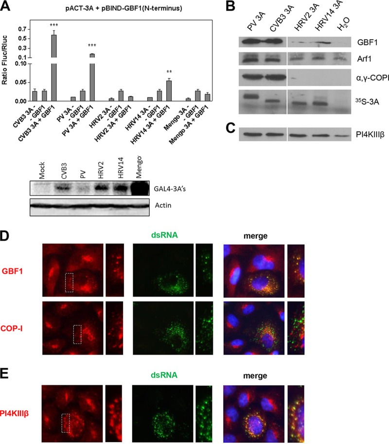
FIG 1.
Interaction of HRV 3A with GBF1. (A) Upper half: interaction of different picornavirus 3A proteins with the N terminus of GBF1 in the mammalian two-hybrid system (M2H). Bars show the means of the results from three samples with standard deviations (SD). Significant differences compared to the highest control determined with a Student's t test are indicated as follows: ** = P < 0.01; *** = P < 0.001. Lower half: Western blotting of picornaviral GAL4-3A proteins tested in M2H. Mengo, mengovirus. (B and C) In vitro HeLa S10 cell extract assay. RNA coding for various enterovirus 3A proteins was translated in HeLa S10 cell extracts. Membranes were isolated by centrifugation and subjected to Western blot analysis to detect the membrane-associated proteins using antibodies against GBF1, Arf1, and α,γ-COPI (B) or PI4KIIIβ (C). The efficiency of the translation reaction was assessed by [35S]methionine labeling. (D and E) BGM cells were infected with HRV2 for 8 h, followed by staining of dsRNA, an infection marker, together with GBF1 or COP-I (D) or PI4KIIIβ (E). Nuclei were visualized with DAPI (4′,6-diamidino-2-phenylindole). The dashed areas are enlarged on the right to show the overlap between signals.

