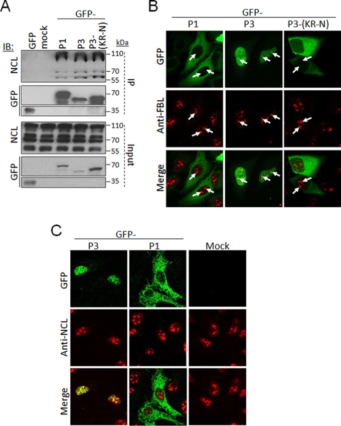
FIG 2.

P-protein can interact with NCL and localize to nucleoli. (A) HEK293T cells were transfected to express the indicated GFP-fused P-proteins or GFP alone or mock transfected, using Lipofectamine 2000. At 18 h posttransfection, cells were lysed, and immunoprecipitation (IP) of GFP-fused proteins was performed using the GFP-trap system (ChromoTek) (8). Lysates (input) and IPs were analyzed by immunoblotting (IB) using antibodies against NCL or GFP. (B and C) HeLa cells transfected to express the indicated proteins were fixed and immunostained with anti-FBL (B) or anti-NCL (C) antibody (Abcam) followed by Alexa Fluor-568-conjugated secondary antibody, and analyzed by CLSM using an Olympus FV1000 microscope with 100× oil immersion objective. The white arrows in FBL-stained samples indicate nucleoli.
