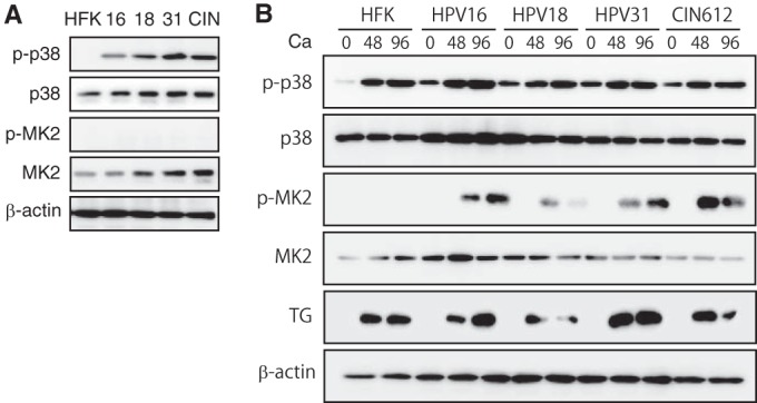
FIG 1.

p-38 and p-MK2 levels are increased in HPV-positive cells. (A) Western blot analysis for p-p38, p38, p-MK2, and MK2 in undifferentiated monolayer cultures of normal human foreskin keratinocytes (HFK), HPV-16, HPV-18, HPV-31, and CIN-612 cells. The HPV-16, -18, and -31 cells were generated by transfection of HFKs with recircularized HPV genomes as previously described (11, 27), followed by selection for a cotransfected drug resistance marker. β-Actin was used as a loading control. The following antibodies were used: p-p38(T180/Y182, D3F9; catalog number 4511), p38(D13E1; catalog number 8690), p-MK2(T334, 27B7; catalog number 3007), and MK2 (catalog number 3042) (all from Cell Signaling Technologies, San Diego, CA). Secondary antibodies included horseradish peroxidase-linked anti-rabbit (Santa Cruz Biotechnology, Santa Cruz, CA). Levels of p-p38, p38, p-MK2, and MK2 following differentiation in high-calcium medium for 0, 48, and 96 h were determined by Western blotting. Calcium-induced differentiation was performed as described previously (27). The 0-h data represent results for undifferentiated cells. TG (transglutaminase) was used as a differentiation marker. β-Actin was used as a loading control.
