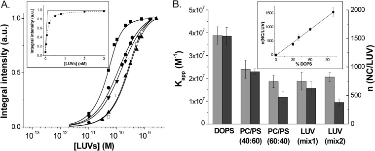
FIG 2.
Determination of the binding parameters of NC protein to model lipid membranes. (A) Titration of MFL-NC (0.1 μM) with LUVs of different lipid compositions: DOPS (■), DOPC-DOPS (40:60) (●), DOPC-DOPS (60:40) (▲), DOPC-DOPE-DOPS-SM-PI(4,5)P2 (16:46:25:8:5) (LUVmix1, ▼), and DOPC-DOPE-DOPS-SM (16:46:30:8) (LUVmix2, □). Solid lines correspond to the fit of the data points with equation 2, using the binding constant and the number of binding sites per LUVs indicated in panel B. (Inset) Determination of the number of binding sites from the intercept of the tangent to the first points of the titration with the tangent to the fluorescence plateau. (B) Binding affinity (light gray) and number of binding sites per LUV (dark gray) as a function of the LUV composition. The values of Kapp and n were determined from the binding curves in panel A. (Inset) Linear dependence of the number of binding sites as a function of the percentage of negatively charged lipids in the LUV composition. All experiments were in 20 mM phosphate buffer, 150 mM NaCl, pH 7.4. Excitation wavelength was 400 nm.

