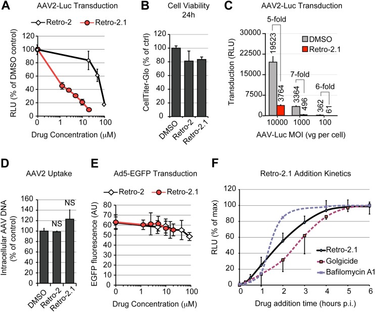
FIG 7.
Inhibitors of STX5 decrease AAV2 transduction and retrograde transport. (A) AAV2-Luc transduction of HeLa cells treated with various concentrations of Retro-2 or Retro-2.1. Cells were pretreated for 30 min at 37°C before addition of AAV2-Luc at an MOI of 10,000 vg/cell. Luciferase activity values are normalized to those for the dimethyl sulfoxide-treated control. (B) Retro-2 and Retro-2.1 are not cytotoxic. HeLa cells were treated with the maximal tested concentrations of Retro-2 (100 μM) and Retro-2.1 (20 μM) for 24 h, and cell viability was measured using the luciferase-based CellTiter-Glo method. (C) AAV2-Luc transduction of HeLa cells at various MOIs in the presence of 10 μM Retro-2.1. Absolute luciferase output and inhibition ratios are indicated. (D) Endocytosis of AAV2-Luc is not reduced in HeLa cells treated with 100 μM Retro-2 or 20 μM Retro-2.1. Intracellular viral DNA was quantified by real-time PCR, and values are normalized to those for the dimethyl sulfoxide-treated control. NS, not significant. (E) Transduction of HeLa cells by Ad5-EGFP in the presence of Retro-2 or Retro-2.1. Values indicate the average ± SD of the green fluorescence intensity per microscope field, expressed in arbitrary units (AU). (F) Kinetics of Retro-2.1 addition. HeLa cells were incubated for 1 h on ice with AAV2-Luc, washed, and transferred to 37°C to trigger infection. Drug was added at various time points, and luciferase activity was measured after 24 h. Dotted lines, the kinetics of golgicide and bafilomycin A1 addition presented in Fig. 1H. Luciferase activity values were normalized to the maximum (plateau) values, as described in the legend of Fig. 1. The values in all panels represent mean and SD from triplicate experiments.

