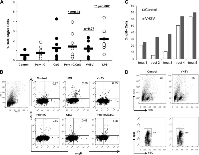
FIG 9.
Effect of VHSV on IgM+ cell proliferation and survival. Spleen leukocytes were incubated with poly(I·C) (50 μg/ml), CpG (5 μg/ml), poly(I·C) combined with CpG, VHSV (1 × 10⁶ TCID50 ml−1), or LPS (100 μg/ml) for 3 days at 20°C. After this time, cells were labeled with BrdU and incubated for a further 24 h. The percentage of proliferating (BrdU-positive) IgM+ cells was then determined as described in Materials and Methods. (A) Percentage of proliferating IgM+ cells out of all IgM+ cells. Circles indicate percentages observed in individual fish, while black bars represent mean values for each group. Significant differences between cells treated with a TLR agonist and control cells are indicated (*, P < 0.05; **, P < 0.01). (B) Dot plots from a representative fish under each condition. (C and D) Spleen leukocytes were infected in vitro with VHSV (1 × 10⁶ TCID50 ml−1), and after 48 h, the number of IgM+ cells in the cultures was determined by flow cytometry. Data shown are percentages of IgM+ cells in control and infected cultures from 5 individual trout, along with dot plots from a representative trout.

