
FIG 8.
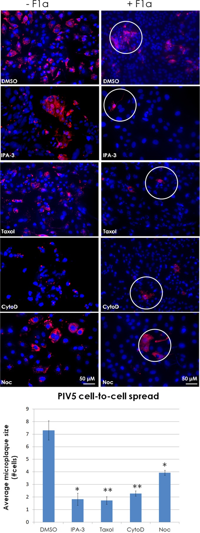
Intercellular connections are utilized by the paramyxovirus parainfluenza virus 5 as a means of cell-to-cell spread. (A) MDCK cells were infected at an MOI of 0.1 with PIV5. At 2 h p.i., cells were treated with neutralizing MAb F1a or left untreated. Cells were then treated with or without IPA-3 (30 μM), paclitaxel (“Taxol”; 100 μM), cytochalasin D (CytoD; 20 μM), or nocodazole (Noc; 30 μM) and incubated for 48 h. Cells were fixed and immunostained for N protein (red), and nuclei were stained with DAPI (blue). Images were taken on a fluorescence microscope with a 20× objective. Microplaque (N-positive neighboring cells, white circles) quantification is shown in the bar graph. *, P < 0.05; **, P < 0.01. Counts for 70 fields of view were included per sample per trial.