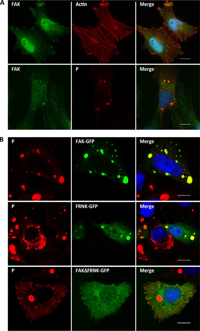
FIG 4.
Localization of FAK in infected cells. (A) Cells were mock infected (top row) or infected with RABV (strain CVS) at an MOI of 3 for 24 h (bottom row). The cells were analyzed by confocal microscopy after staining with anti-P antibody, rabbit anti-FAK antibody, or Texas red-phalloidin. DAPI (blue) was used to stain the nuclei (Merge). Colocalization is apparent as yellow coloration in the merged images. Scale bars, 15 μm. (B) Cells were transfected with plasmids encoding FAK-GFP, FRNK-GFP, or FAKΔFRNK-GFP and then infected at an MOI of 3 for 24 h. The cells were analyzed by confocal microscopy after staining with anti-P antibody. DAPI (blue) was used to stain the nuclei (Merge). Colocalization is apparent as yellow coloration in the merged images. Scale bars, 15 μm.

