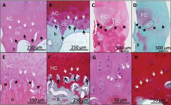
Figure 1.

Different features of the osteochondral junction in normal and repair cartilage are revealed by hematoxylin and eosin (H & E) (A, C, E, G) and Safranin O/fast green/iron hematoxylin (SafO) (B, D, F, H). In normal human cartilage (A and B, adult hip surgical waste, femoral neck fracture), H&E clearly stains the tidemark (A, white arrows), while SafO readily discriminates cartilage from fast green–stained bone (below the black arrows, B). For heterogeneous human repair cartilage (C and D, biopsy taken 1 year postmicrofracture71,121), H&E is better for determining the cartilage-bone boundary (black arrows, C) and abnormal mineralization (dashed circle), while SafO discriminates fibrocartilage from fast green–stained fibrous repair and bone (D). In hyaline cartilage repair elicited in a sheep model (E-H, 6 months posttreatment43), the tidemark is beginning to form (white arrows, 10x magnification for E and F, 40x magnification for G and H). White arrows = tidemark; black arrows = cartilage-bone interface; AC = articular cartilage; cc = calcified cartilage; FC = fibrocartilage; HC = hyaline cartilage; b = bone.
