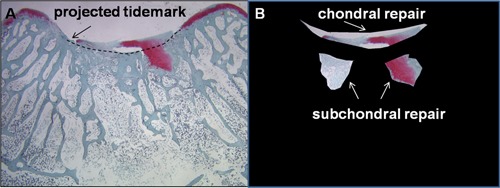
Figure 5.

Histomorphometry of chondral versus subchondral soft repair tissues. The example is from a 2-month repair of a trochlear full-thickness rabbit knee defect with two 0.9-mm microdrill holes.44 (A) Safranin O–stained trochlear repair tissue, with the “projected tidemark” drawn through the defect area. (B) The chondral repair is cropped separately from the subchondral soft tissue repair for further histomorphometric analysis.
