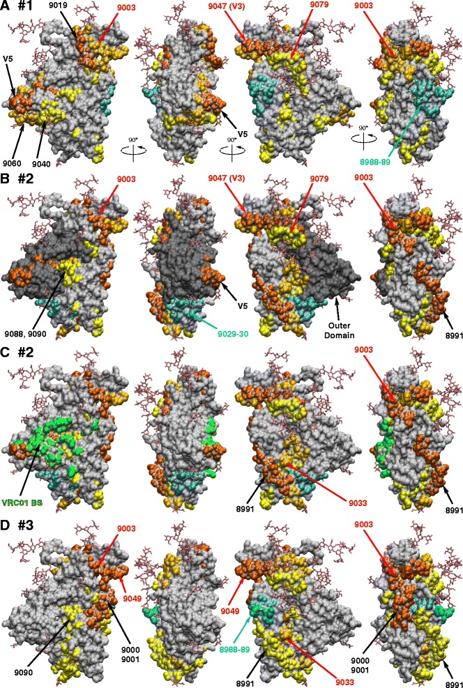
Figure 2.

Immunogenic linear epitopes plotted onto the surface of gp120. ELISA data from Figure 1 were used to illustrate immunogenic epitopes for rabbits #1 (A), #2 (B and C), and #3 (D). A450 reading between 1–2, 2–3, and 3–4 are shown in yellow, gold and orange, respectively. Peptides with reduced antibody reactivity after the second immunization are shown in cyan. Peptides that were reactive in all three animals are indicated in red text. Outer domain is shown in panel B in dark grey and VRC01 binding site is shown in green in panel C. The crystal structure of SOSIP gp140 (accession code 4NCO) was used to generate figures.
