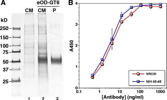
Figure 4.

Expression, purification and antigenic analysis of eOD-GT6. (A) Expression and purification of eOD-GT6. Protein was detected by silver staining. Lane 1: culture medium (CM) of mock transfected cells; lane 2: CM of eOD-GT6 transfected cells, 4 days after transfection; lane 3: purified (P) eOD-GT6. (B) eOD-GT6 ELISA with VRC01 (red) and NIH45-46 (blue).
