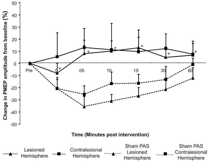
Figure 2.
Group mean percentage of change in PMEP amplitude after PAS10min over the lesioned hemisphere and after PASSham (protocol 4). A significant increase in amplitude is observed ipsilaterally after PAS10min applied to the virtually focal suppressed stronger pharyngeal representation (*P < .005) (▲). However, there was no significant increase in cortical excitability of the contralesional hemisphere (■). Hemispheric responses (lesioned and contralesional) were compared with the responses of each hemisphere after PASSham (dashed lines).

