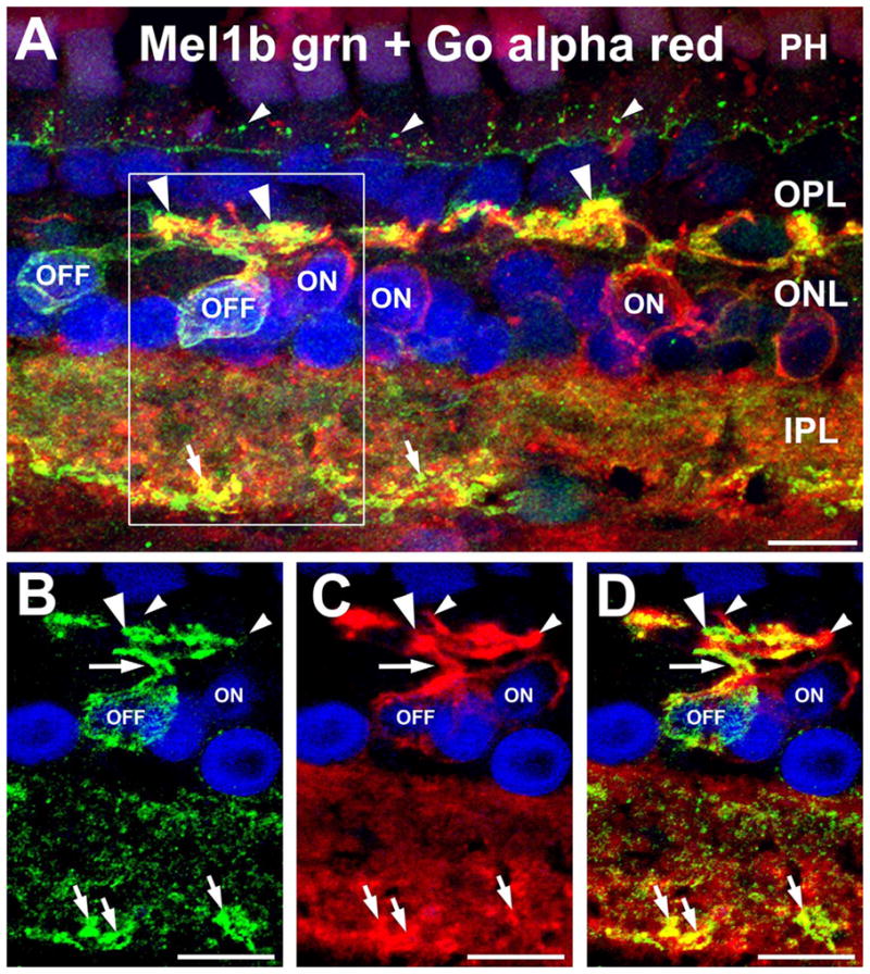
Figure 4.

Mel1b receptors are expressed by OFF bipolar cells. A: Double labeling for Mel1b receptors (green) and the ON bipolar cell marker Goα (red) shows that Mel1b receptor-immunoreactivity is absent from the cell bodies of ON bipolar cells (ON), identifying the Mel1b receptor-immunoreactive bipolar cells as OFF bipolar cells (OFF). Confocal image stack comprised of seven optical slices of 400 nm each. Apparent colocalization of Mel1b and Goα immunoreactivity in processes in the outer plexiform layer (OPL) is due to their close proximity and the relative thickness of the image stack, and does not represent genuine colocalization (see panels B–D, below). Immuno-labeling for both Mel1b and Goα is present in the inner plexiform layer (IPL), with strongly Mel1b-positive processes (small arrows) present along the inner margin of the layer. Mel1b immunoreactive puncta (small arrowheads) are also present at the level of the outer limiting membrane (OLM). The box in (A) indicates area shown in panels B–D. B–D: Examination of a single confocal optical plane confirms that Mel1b labeling does not colocalize with labeling for Goα in ON bipolar cells. Images in panels B–D represent a single optical slice of ≈400 nm. B: Mel1b immunoreactivity in the cell body and primary and secondary dendrites (arrow and large arrowhead respectively) of a bipolar cell (OFF). C: ON bipolar cell dendrites labeled for Goα (small arrowheads). D: Overlay of panels B,C showing that ON bipolar cell dendrites are devoid of Mel1b receptor labeling. Nuclei are counterstained with DAPI (blue) in all panels. INL, inner nuclear layer; PH, photoreceptor inner segments. Scale bars = 10 μm in all panels.
