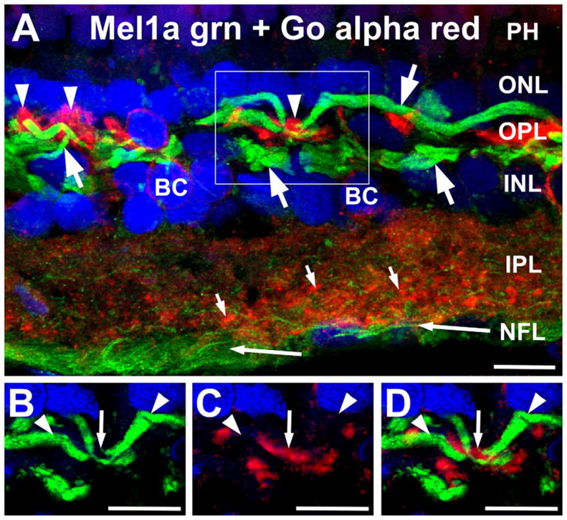
Figure 6.

Mel1a receptors are not expressed in ON bipolar cell dendrites. A: Labeling for Mel1a receptor (green) and Goα (red) localize to different processes in the outer plexiform layer (OPL). Mel1a receptors are expressed in distinctive processes morphologically similar to horizontal cell axons (large arrows). These processes are distinct from the Goα-immunoreactive ON bipolar cell dendrites (arrowheads). Apparent colocalization of Mel1b and Goα immunoreactivity in processes in the outer plexiform layer (OPL) is due to their close proximity and the relative thickness of the image stack, and does not represent genuine colocalization (see panels B–D, below). Mel1a immunoreactivity also is present in puncta throughout the inner plexiform layer (IPL; small arrows) and in ganglion cell axons (long arrows) in the nerve fiber layer (NFL). Confocal image stack comprised of 15 optical slices of ≈400 nm each. The box indicates the area shown in panels B–D. B–D: Examination of thin stacks of confocal optical planes confirms that Mel1a labeling is not localized to ON bipolar cell dendrites. Images in panels B–D are comprised of five optical slices of ≈400 nm each. B: Mel1a immunoreactivity in horizontal cell axons (arrow-heads) in the OPL. C: ON bipolar cell dendrites labeled for Goα (small arrow). D: Overlay of panels B,C showing that ON bipolar cell dendrites (Goα-positive) and Mel1a receptor-positive processes are not colocalized. Nuclei are counterstained with DAPI (blue) in all panels. BC, Bipolar cell; PH, photoreceptors; IPL, inner plexiform layer; NFL, nerve fiber layer. Scale bars = 10 μm in all panels.
