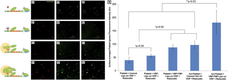
Fig. 6.

(A1–D3) Representative fluorescence microscopy images (along with envisioned mechanistic schema) of peptide-decorated rhodamine-labeled (red) constructs and calcein-stained (green) platelets incubated simultaneously on Risto-treated VWF adsorbed on glass coverslips. (E) Quantitative overall fluorescence intensity data of platelets (green) adhered and aggregated on the VWF-adsorbed coverslips. A1, B1, C1 and D1 represent construct binding; A2, B2, C2 and D2 represent platelet binding; A3, B3, C3 and D3 represent merged results to exhibit co-localization in yellow. The conditions tested were undecorated (Unmod-Lipo), VBP-decorated (VBP-Lipo) and VBP–FMP-co-decorated (VBP–FMP-Lipo) liposomal nanoconstructs incubated with predominantly inactive platelets (Platelet) and ADP-activated platelets (Act Platelet). Undecorated constructs showed minimal VWF-binding and platelet co-localization, VBP-decorated constructs showed concomitant VWF-binding with platelets but minimal platelet co-localization, and VBP–FMP-co-decorated constructs showed substantial VWF-binding as well as platelet co-localization, especially if platelets were already in a pre-activated state.
