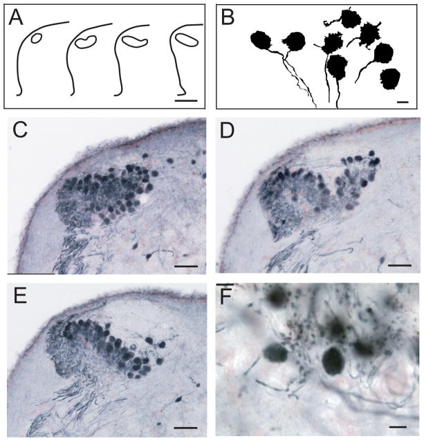
Figure 3.
Organization and cellular architecture of NM, after injection of NB in the contralateral NL. A: Outline drawings of NM in transverse sections at 100-μm intervals from caudal (left) to rostral. B: Drawing of labeled neurons with large, round, rugose cell bodies and ventrally orientated NM axons. C: Profile of caudal NM with most neurons labeled at the dorsolateral region. D: Neurons in middle NM. E: Neurons in rostral NM; note their altered dorsomedial-ventrolateral orientation. F: High-power photomicrograph showing labeled neurons in NM with round or ellipsoid form. Scale bars = 200 μm in A; 10 μm in B; 50 μm in C–E; 15 μm in F. [Color figure can be viewed in the online issue, which is available at wileyonlinelibrary.com.]

