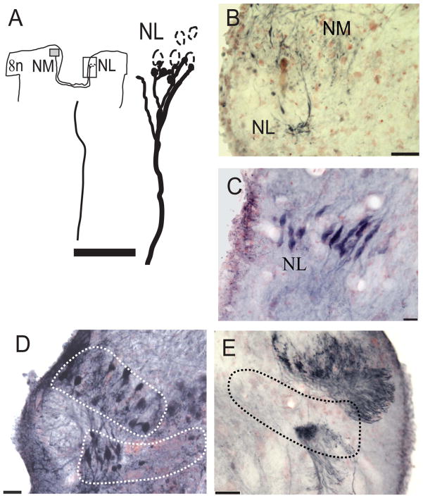
Figure 4.
Connections and cellular architecture of NL labeled by injection of NB to NM/NL. A: Drawing to show an injection site in NM (gray box) and NM axons that terminated in contralateral ventral NL. An enlarged view of the rectangular frame surrounding the contralateral NL shows a single well-labeled axon, which formed terminal boutons on the ventral dendrites of NL neurons. B: Terminals in NL were labeled with a small injection of NB in the ipsilateral NM. C: Bitufted neurons labeled after a small injection of NB into the fiber tract entering the torus semicircularis. D: NB injection site in NM and NL revealed round cell bodies in NM and bitufted cells in NL. E: The contralateral side of the same case as in D, showing retrogradely labeled neurons in NM and fibers in ventral NL. Scale bars = 50 μm in A,B,E; 20 μm in C,D. [Color figure can be viewed in the online issue, which is available at wileyonlinelibrary.com.]

