
Figure 8.
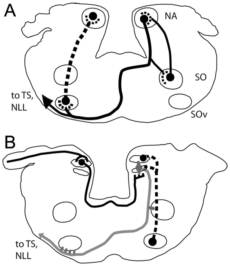
Summary of the connections of the cochlear nuclei and superior olive complex. A: NA innervated mainly the ipsilateral lateral portion of SOd, the contralateral SOv, and the contralateral torus semicircularis. SOd and SOv both projected back to the ipsilateral first-order nuclei. B: NM projected to the dorsal neuropil of the ipsilateral NL and across the midline to the ventral neuropil of the contralateral NL, whereas NL projected to the ipsilateral SOd and to the auditory midbrain, through a fiber bundle that descended to run ventral to the contralateral SOv (gray line). SOv projected back to the ipsilateral NM and NL (dashed line).