
Figure 3.
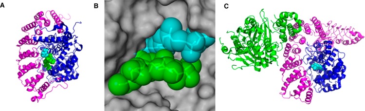
Crystal structures of prenyltransferase enzymes. (A) Crystal structure of FTase in complex with a nonhydrolyzable FPP analog and a peptide substrate based on KRas-4B (PDB 1D8D): magenta, α-subunit; blue, β-subunit; cyan, isoprenoid analog; green, CaaX peptide. (B) Binding pocket of FPP showing interaction of protein and isoprenoid substrates over a large surface area: gray, space-fill structure of β-subunit of FTase; cyan, isoprenoid analog; green, CaaX peptide. (C) Crystal structure of GGTase-II in complex with Rab escort protein and FPP (PDB 1LTX): magenta, GGTase-II α-subunit; blue, GGTase-II β-subunit; green, Rab escort protein; cyan, FPP.